
AI Photo Editing for Myrmecologists — Magic Eraser
How myrmecologists use AI photo editing for ant specimen records, taxonomic photography, and research publications. Remove pin artifacts, enhance morphological details, and create publication-ready figure plates for ant systematics.

SEO & Growth
Reviewed by Magic Eraser Editorial ·
Table of Contents
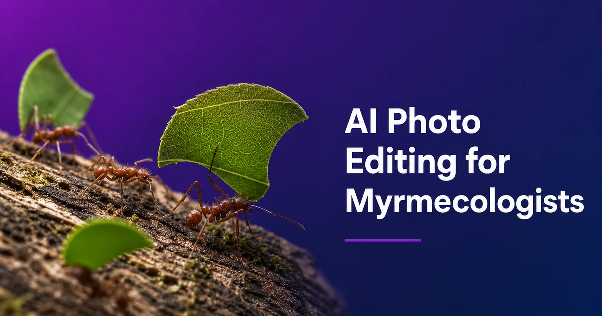
Myrmecology — the scientific study of ants — depends on high-quality specimen photography spanning scales from field habitat records to individual body structures measured in hundredths of millimeters. With over 16,000 described ant species and an estimated 6,000 to 8,000 species still awaiting formal description, the demand for clear, standardized specimen imaging has never been greater. Every new species description requires detailed figure plates showing diagnostic morphological features from multiple standardized views, and the revolution in digital taxonomy databases. Mainly AntWeb, which now hosts images of over 90,000 ant specimens — has made photographic records as fundamental to ant systematics as the physical specimens themselves.
The photographic challenges in myrmecology are driven by scale, standardization needs, and the sheer diversity of material that must be imaged. Most ant workers measure between two and fifteen millimeters in length, placing them in the technically demanding range where high-magnification macro photography is necessary but the specimens are large enough to have complex three-dimensional form. A single worker ant may have sculpturation patterns on the head surface, teeth along the mandible margin, spines projecting from the propodeum, and hair-like setae covering the body. All of which must be clearly resolved and distinguished in photographs that will be examined at extreme zoom by taxonomists worldwide. Focus stacking is key but introduces artifacts. Lighting must be diffused enough to reveal surface sculpture without flattening the three-dimensional form.
AI photo editing tools address these challenges by automating labor-intensive post-processing steps that otherwise consume a substantial fraction of myrmecological research time. Pin and mount removal produces clean specimen images suitable for databases and publications. Detail boost recovers the fine morphological structures — mandible dentition, sculpturation patterns, pilosity traits — that determine species identification. Color normalization compensates for the dramatic variation in specimen look caused by different preservation methods, ages, and photographic conditions. For myrmecologists managing field collections, museum visits, teaching loads, and publication deadlines at once, efficient AI-assisted image processing directly accelerates the pace of taxonomic research.
- Pin, point-mount, and background removal produces clean specimen images meeting the standardized imaging protocols of AntWeb and other digital taxonomy databases.
- AI boost sharpens diagnostically critical morphological structures — mandible dentition, propodeal spines, petiole shape, sculpturation, and pilosity — at the magnification levels taxonomy requires.
- Color normalization compensates for preservation artifacts, age-related darkening, and variable lighting across specimens photographed at different institutions and time periods.
- Focus stacking artifact cleanup eliminates halos, edge doubling, and fringing that compromise the clarity of high-magnification composite images.
- Multi-view figure plates in dorsal, lateral, and full-face orientations are exported at 300 DPI for journal submission and taxonomic database contribution.
Multi-view standardized imaging and AI-assisted post-processing
Ant taxonomic photography follows strict standardization protocols developed over decades to ensure that images from different researchers, institutions, and equipment can be meaningfully compared. The three required views — dorsal (looking down on the specimen from directly above), lateral (profile view from the side at specimen midline). Full-face (head viewed from directly in front) — each reveal different diagnostic characters. The dorsal view shows head shape, mesosoma outline, and the relative proportions of body segments. The lateral view reveals propodeal spines, petiole and postpetiole profiles, and the overall body architecture. The full-face view exposes mandible shape, clypeal structure, eye position, and antennal insertion points. All three views must be captured with the specimen in a precisely controlled orientation. Requires specialized staging equipment and considerable patience.
AI post-processing integrates into each view's workflow at multiple points. After focus stacking merges the 30 to 80 frames captured per view into a single all-in-focus composite, the AI removes the stacking artifacts that invariably appear. Halos around high-contrast edges, doubled edges from slight alignment errors between frames, and fringing around projecting structures like spines and antennae that move slightly between exposures on older specimens with loosened joints. Pin removal is the most universal cleanup operation: every pinned specimen shows the stainless steel pin passing through the right side of the mesosoma, often with visible adhesive. This must be removed for publication-quality images while keeping the mesosoma surface it penetrates.
Background standardization is critical for database contribution. AntWeb and similar platforms display specimen images on plain white backgrounds with consistent lighting quality. Images submitted with cluttered or inconsistent backgrounds are either rejected or require time-consuming manual editing. The AI removes not just the physical background of the photography setup but also the mounting substrate. Pin staging platforms, point-mount paper triangles, and cotton wool used in some museum preparations — producing isolated specimen images that meet database standards without the hours of manual masking that these complex, irregular shapes would otherwise require.
- Three standardized views — dorsal, lateral, and full-face — each reveal different diagnostic characters essential for species-level identification in ant taxonomy.
- Focus stacking of 30 to 80 frames per view introduces artifacts that AI cleanly eliminates — halos, edge doubling, and fringing around projecting spines and antennae.
- Pin removal through the mesosoma is the most universal cleanup task, preserving the body surface around the pin insertion while erasing the steel shaft and adhesive.
- Background standardization to plain white meets AntWeb and database submission protocols that require consistent imaging standards across all contributed specimens.
Enhancing diagnostic morphological characters for ant identification
Ant species identification relies on a precise hierarchy of morphological characters that photographs must clearly resolve for scientific utility. At the genus level, mandible form. The number, size, and arrangement of teeth along the masticatory margin — is often the primary diagnostic feature, with some genera defined by two teeth and others by twelve or more. Clypeal structure, the form of the frontal carinae, and the number of antennal segments further distinguish genera. At the species level, finer characters take precedence: the specific shape and length of propodeal spines, the profile of the petiole and postpetiole nodes, the density and pattern of surface sculpturation (whether rugose, punctate, reticulate, or smooth). The type, density, and distribution of body hair (pilosity and pubescence). AI boost increases local contrast and sharpness to make each of these character systems clearly legible in photographs.
Surface sculpturation is mainly challenging to photograph because it exists as very shallow three-dimensional texture on the body surface. Rugae — the raised ridges that cover the head and mesosoma of many ant genera — may stand only a few micrometers above the surrounding surface. Their visibility depends fully on the angle and quality of lighting relative to the surface. Punctures — tiny circular depressions scattered across the cuticle — are even more subtle. AI boost compensates for imperfect lighting by increasing the micro-contrast that makes these surface features visible, recovering sculpturation detail in areas where the original lighting angle happened to be parallel to the surface and rendered the texture invisible.
Pilosity — the body hair system — is one of the most diagnostically important and photographically difficult character systems in ant taxonomy. Ants bear multiple types of body hair: erect setae that project perpendicular to the body surface, decumbent or appressed pubescence that lies flat against the cuticle. Specialized hair forms on specific body regions. The distinction between these hair types, their density, their distribution pattern. Their precise form (straight, curved, branched, or clavate) separates closely related species. These structures are often translucent and very fine. A single seta may be only a few micrometers in diameter — making them difficult to photograph clearly and nearly invisible against anything but a perfectly clean, contrasting background.
- Mandible tooth count and arrangement is often the primary genus-level diagnostic character — AI enhancement must resolve individual teeth along the masticatory margin.
- Surface sculpturation stands only micrometers above the cuticle surface; AI micro-contrast enhancement recovers detail where original lighting angle rendered texture invisible.
- Propodeal spine shape and length, petiole profiles, and postpetiole form are species-level characters that require sharp edge definition in lateral view photographs.
- Pilosity types — erect setae, appressed pubescence, and specialized regional hairs — are translucent and micrometer-thin, requiring clean backgrounds and maximum image clarity.
Processing museum collections and managing preservation artifacts
The world's ant collections — estimated at over 100 million specimens in natural history museums and research institutions globally — represent an irreplaceable biodiversity archive accumulated over more than 250 years of collection effort. These specimens are the physical evidence underlying every published species name, and type specimens. The specific specimens that serve as the reference standard for each species name — are the most scientifically valuable objects in any collection. Photographing type specimens for digital databases has become a priority for major museums, but the condition of historical material presents consistent photographic challenges: old adhesives, corroded pins, faded labels obscuring body parts. The physical deterioration of specimens that may be over a century old.
AI processing tools maximize the information extractable from museum material without physical manipulation that risks damaging irreplaceable specimens. Boost applied to photographs of older specimens increases the visibility of surface features obscured by age-related changes. Cuticular darkening, the accumulation of dust and adhesive residue, and the general loss of surface contrast that occurs as specimens oxidize over decades. Pin removal is mainly valuable for historical specimens where the pin is corroded, bent, or has deposited verdigris staining onto the specimen surface. The AI removes the pin artifact while keeping or even enhancing the visibility of the morphological features right away surrounding the pin insertion point.
Specimen photography campaigns at major museums may involve imaging thousands of specimens during a single research visit, under time pressure with whatever lighting equipment is available at the host institution. AI batch processing standardizes the results of these intensive imaging sessions, applying consistent background removal, color correction. Boost across hundreds of images captured under variable conditions. This batch capability transforms the economics of museum records. A visiting researcher can focus on capturing maximum coverage rather than spending time perfecting individual images in the field, confident that AI post-processing will bring the entire set to publication standard.
- Over 100 million ant specimens in global collections provide the physical evidence for every published species name — type specimens are scientifically irreplaceable.
- AI enhancement recovers surface features obscured by age-related darkening, adhesive residue, dust accumulation, and the general contrast loss of oxidized cuticle.
- Corroded or verdigris-stained pins on historical specimens are removed while preserving morphological detail around the insertion point.
- Batch processing standardizes imaging from intensive museum visits where thousands of specimens are captured under variable conditions and time pressure.
Ecological surveys, citizen science, and educational outreach
Ant ecology increasingly relies on photographic spotting for large-scale biodiversity surveys where collecting every specimen is impractical, undesirable, or prohibited. Camera traps, smartphone macro photography, and standardized photo-sampling protocols generate thousands of images per study that must be sorted and identified. And while AI-based automated species identification is advancing rapidly, the quality of input images at its core limits identification accuracy. AI photo boost applied to field-collected images before they enter spotting pipelines improves the resolution of diagnostic features, increasing the proportion of images that can be identified to species level rather than stopping at genus or subfamily. For ecological studies tracking community composition, the difference between species-level and genus-level spotting can determine whether meaningful ecological conclusions are possible.
Citizen science platforms like iNaturalist receive hundreds of thousands of ant observations annually. The spotting rate for ant photos is substantially lower than for more photogenic insect groups. Most casual observers photograph ants from above with a smartphone, producing dorsal-view images that lack the magnification needed to see diagnostic characters like mandible dentition or propodeal spines. AI boost can sharpen the characters that are visible. Head shape, relative body proportions, color pattern — to the point where experienced identifiers can place the observation to genus level, which is enough for most distribution mapping and biodiversity monitoring purposes. Improving the photographic quality of citizen science ant observations expands the effective survey network for myrmecological conservation without requiring every contributor to own specialized macro equipment.
Educational content about ants benefits enormously from high-quality specimen photography because the animals' most remarkable features. Polymorphic caste systems, specialized mandible forms for different feeding strategies, the structural complexity of the mesosoma with its fused thoracic segments — are all invisible at the scale of casual observation. AI-enhanced macro and focus-stacked photography transforms working research images into visually strong content that reveals ant morphological complexity to general audiences. These enhanced images serve museum exhibits, university entomology courses, science communication publications. The growing public interest in ant biology driven by popular science content creators. Making the microscopic world of ant anatomy visible builds support for biodiversity conservation by connecting people with the complexity of organisms they encounter daily but never truly see.
- AI enhancement of field survey images increases the proportion identifiable to species level — critical for ecological studies tracking community composition.
- Citizen science ant observations on iNaturalist gain identification value when AI sharpening resolves head shape, proportions, and color patterns visible in casual smartphone photos.
- Batch enhancement of camera-trap and photo-sampling images makes large-scale ecological survey datasets practical to process within research timelines.
- Educational outreach uses AI-enhanced macro photography to reveal ant morphological complexity — caste polymorphism, mandible diversity, and mesosoma architecture — to general audiences.
How to do it
- 1
Photograph ant specimens using standardized multi-view macro setups
Set up a macro photography station with diffused lighting for the three standard taxonomic views required in ant systematics. Dorsal (top), lateral (side), and full-face (head-on). Include a millimeter scale bar in every frame. For pinned museum specimens, use a motorized focus rail to capture focus stacks of 30 to 80 frames per view at high magnification. Individual worker ants range from one to thirty millimeters in length and depth of field at the necessary magnification covers only a fraction of the specimen. For field photography, photograph living ants in controlled settings or freshly collected material before preservation alters color and posture.
- 2
Remove pin artifacts, mounting debris, and background clutter
Use Magic Eraser to remove the insect pin, staging platform, point-mount card or glue residue. Any background elements that distract from the specimen. Pinned ant specimens always show the pin passing through the mesosoma, often with visible glue or shellac around the insertion point. Point-mounted specimens are glued to a small paper triangle, which partially obscures ventral characters. Background removal produces clean specimen images on white or neutral backgrounds that meet the standards of digital taxonomy databases like AntWeb. Standardized imaging has been central to the platform since its founding.
- 3
Enhance fine morphological structures critical for species identification
Use AI Enhance to sharpen the specific body structures that drive ant identification at the genus and species level. Mandible tooth count and shape, clypeal structure, antennal segment proportions, propodeal spine length and form, petiole and postpetiole shape, sculpturation patterns on the head and mesosoma, and the density and arrangement of body pilosity. Many of these characters are measured in tenths of millimeters. Their clear resolution in photographs is key for species descriptions, spotting keys, and the growing use of image-based spotting in ecological surveys.
- 4
Correct color and lighting inconsistencies across specimen series
Use AI color correction to standardize look across series of specimens photographed under different conditions. Fresh collections versus decades-old museum material, different lighting equipment at different institutions, and the natural color variation between preserved and living specimens. Ants preserved in ethanol often lose their natural coloration, turning uniformly reddish-brown or yellow. Dry-mounted specimens darken over time. Consistent color normalization across a publication's figure plates ensures that readers can meaningfully compare the morphology shown in different images without being misled by keeping or photographic artifacts.
- 5
Prepare multi-view figure plates for taxonomic publications and databases
Arrange the standardized dorsal, lateral, and full-face views into publication-ready figure plates with consistent spacing, scale bars, and specimen labels. Export at 300 DPI minimum for journal submission to taxonomic publications like Zootaxa, Myrmecological News, or the Journal of Hymenoptera Research. For AntWeb and similar databases, prepare extra web-optimized versions at standardized dimensions. For spotting guides and citizen science platforms, create annotated versions with key diagnostic features labeled to bridge the gap between what non-specialists photograph in the field and the precise morphological characters that expert spotting requires.
Sources
- Photographic Standards for Ant Taxonomy: Specimen Imaging Protocols — AntWeb — California Academy of Sciences
- Morphological Character Systems in Formicidae: An Atlas of Ant Body Structures — AntWiki — Global Ant Biodiversity Informatics
- Focus Stacking Techniques for Insect Macrophotography and Taxonomic Documentation — ZooKeys — Pensoft Publishers