
AI Photo Editing for Mycologists: Document Fungi Specimens — Magic Eraser
Expert fungi photography editing for mycologists and mushroom researchers. AI-powered tools for specimen records, spore print photography, gill detail boost, and field identification imagery.
SEO & Growth
Reviewed by Magic Eraser Editorial ·
Table of Contents
- Field photography technique: capturing diagnostic features for reliable identification
- Background removal and specimen isolation for taxonomic documentation
- Enhancing diagnostic features: gill detail, surface texture, and color fidelity
- Specialized applications: spore prints, microscopy, and citizen science platforms
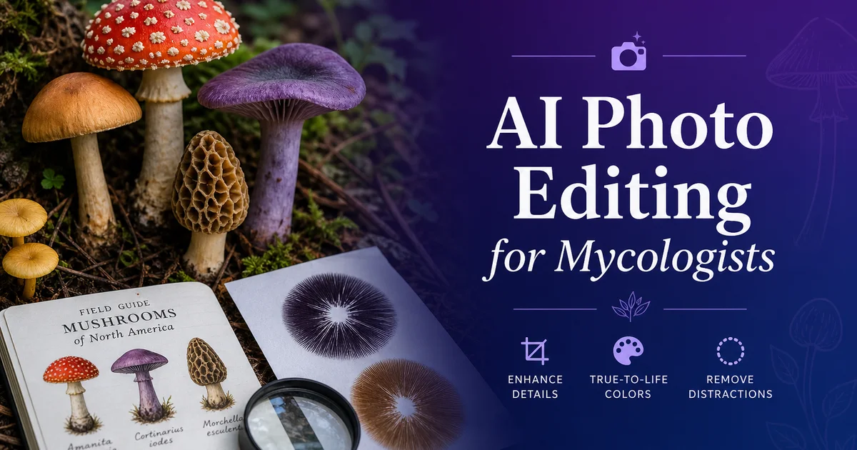
Mycology — the scientific study of fungi — depends on visual records more heavily than almost any other biological discipline, because the morphological features that distinguish one fungal species from another are often subtle surface textures, precise color hues. Three-dimensional structural details that are difficult to describe in words but right away apparent in a well-made photograph. A mycologist examining a mushroom in the field evaluates cap shape and surface texture, gill spacing and attachment type, stipe surface character, veil remnants, spore print color, flesh color and any bruising reactions, odor, habitat, and substrate. And the photograph they take needs to capture enough of these features to support identification review later at a desk or by colleagues who cannot examine the physical specimen. For expert mycologists, citizen science contributors, field guide authors. The growing community of amateur enthusiasts, photography is the bridge between field observation and reliable identification.
The technical challenges of fungi photography are formidable and specific to the subject matter. Mushrooms grow in the most photographically difficult settings imaginable. Deep forest shade, rotting logs in dappled light, leaf litter with complex visual textures, and soil settings where brown specimens blend into brown backgrounds. The specimens themselves present optical challenges that test camera systems: gills are delicate parallel structures that require precise lighting angle to resolve, cap surfaces range from mirror-glossy viscid coatings to matte dry textures that absorb light differently, colors span the entire visible spectrum and often include subtle hues that cameras struggle to reproduce accurately. The three-dimensional form of a mushroom changes the lighting need from one angle to the next. Most field mycologists work with phone cameras under available light, producing images that capture the general gestalt of a specimen but lose the fine detail needed for confident species-level identification.
AI photo editing tools address the specific visual documentation challenges that mycologists face at every stage of their workflow. Background removal isolates specimens from the visually chaotic forest floor, enabling clean display in field guides, identification databases, and research publications. AI Enhance recovers the gill detail, surface textures, and color nuances that camera limitations and field lighting conditions degrade. Object removal cleans up the debris, insect damage, and handling artifacts that are inevitable in field specimen photography. This guide covers the complete mycological photography and editing workflow, from field collection techniques through specialized applications like spore print records and microscopy image preparation, with emphasis on the diagnostic features that make fungi photography scientifically valuable rather than merely decorative.
- Background Eraser isolates fungal specimens from complex forest floor settings, enabling clean taxonomic plates, field guide illustrations, and database-ready images with standardized backgrounds.
- AI Enhance recovers the diagnostic morphological details — gill spacing, cap surface texture, veil remnants, and stipe character — that phone cameras lose in challenging forest shade lighting.
- Color accuracy preservation during enhancement is critical in mycology where subtle hue differences between cap, gill, and flesh can distinguish edible species from dangerous toxic lookalikes.
- Magic Eraser removes field photography complications including soil debris, insect damage, slug trails, leaf fragments, and handling artifacts that obscure diagnostic specimen features.
- Batch export produces format-specific images for taxonomic publications, herbarium databases, citizen science platforms, field guide printing, and educational displays from a single edited master.
Field photography technique: capturing diagnostic features for reliable identification
The foundation of useful mycological photography is a systematic approach to capturing every diagnostic feature a specimen offers. The feature that clinches an identification is often not the one you expect when you press the shutter. Expert mycologists and experienced citizen scientists develop a standardized shooting protocol for each collection: a habitat shot showing the specimen in its growth context and substrate type, a top-down cap view revealing surface texture and color pattern, an underside view showing the hymenium type (gills, pores, teeth, or smooth surface) and its attachment to the stipe, a full profile shot from the side showing overall proportions and any veil remnants. A cross-section cut revealing the flesh color, thickness, and any bruising or color-change reactions that occur upon exposure to air. This multi-angle protocol ensures that identification-critical features are documented even when the photographer does not yet know which features will prove diagnostic for the particular species.
Lighting management in the field is the single most impactful technical factor in fungi photography. The difference between useful and useless specimens often comes down to light angle. The critical principle is that three-dimensional structures like gills, pores, and surface textures become visible through shadow. And shadow requires directional light at an angle to the subject surface. Direct overhead light, whether from midday sun or camera flash, illuminates every surface equally and eliminates the shadows that reveal texture and structure. The most effective field technique is to position yourself so that available light hits the specimen from the side or slightly behind, creating shadows that reveal gill edges, surface bumps, fibrillose textures. The dimensional form of the cap and stipe. A small reflector card — even a white notebook page — bounced into the shadow side prevents the contrast from becoming too extreme for the camera sensor.
Spore print photography is a specialized documentation task that many mycologists find surprisingly difficult to execute well. A spore print is made by placing a mushroom cap gill-side-down on paper (half white, half dark) for several hours and allows the spore deposit color to be assessed. A critical identification feature for many genera. The resulting print is a delicate deposit of microscopic spores that can range from white through cream, pink, brown, rusty-orange, purple-brown, to jet black. Photographing spore prints requires perfectly even lighting with no specular reflections off the paper surface, careful color management to capture the true print hue. Enough resolution to show the print pattern that corresponds to the gill arrangement. Phone cameras tend to blow out white prints to pure white and compress dark prints to featureless black, losing the subtle color information that mycologists need.
- A standardized multi-angle shooting protocol — habitat, cap top, gill underside, profile, and cross-section — ensures all diagnostic features are documented regardless of which proves critical for spotting.
- Directional side lighting is essential for revealing three-dimensional gill structures, surface textures, and veil remnants that disappear under flat overhead illumination or camera flash.
- A white reflector card bounced into the shadow side of a directionally lit specimen prevents excessive contrast that overwhelms phone camera sensors in forest shade conditions.
- Spore print photography requires even shadowless lighting, accurate color reproduction, and enough resolution to capture the subtle hue differences between white, cream, pink, and other light-colored deposits.
Background removal and specimen isolation for taxonomic documentation
The visual complexity of natural fungal habitats is the primary obstacle to clean specimen records. Background Eraser addresses this directly by isolating the specimen from its setting. A mushroom growing from leaf litter on a forest floor exists in a visual field of brown-on-brown-on-brown. Dead leaves, soil, twigs, other decomposing organic matter — that makes it difficult for viewers to distinguish specimen edges from substrate even when they know what they are looking at. The problem intensifies with species that have brown or tan coloring that closely matches their growth substrate, small specimens growing in clusters where individual fruiting bodies overlap. Specimens growing from the underside of logs or branches where the growth substrate itself becomes a confusing visual element. Background removal separates the specimen cleanly from all of this environmental noise.
The choice of replacement background depends on the documentation purpose and should follow established conventions within mycological publishing. Taxonomic plates and formal species descriptions in the past use white or very light gray backgrounds that maximize color accuracy and allow diagnostic features to be compared across plates without background variation. Field guide illustrations often use a simplified natural background. A suggestion of woodland texture without the specific visual complexity of the actual collection site — that provides context without distraction. Digital identification databases and apps benefit from transparent backgrounds that allow specimens to be composited with diagnostic labels, measurement annotations, and comparison images. For each purpose, the AI background replacement produces consistent, standardized results that would require a portable studio setup to achieve in the field.
Keeping the basal mycelium, substrate attachment point, and any rhizomorphs or mycelial cords connected to the specimen base is important when isolating mushroom specimens because these features carry diagnostic information. Some species are identified partly by how their stipe base relates to the soil. Whether it terminates in a bulb, a volva sac, a rooting extension, or spreading mycelium. Background Eraser needs to be applied selectively here, removing the surrounding substrate while keeping the soil or wood material right away attached to the stipe base that reveals the growth habit. This selective approach produces images that are clean and focused while retaining the morphological information at the critical junction between fungal fruiting body and growth substrate.
- Forest floor backgrounds create brown-on-brown visual complexity that makes specimen edges difficult to distinguish, especially with tan or brown species growing on matching substrates.
- Taxonomic plates use white backgrounds for color accuracy, field guides use simplified natural settings for context, and digital keys use transparent backgrounds for annotation compositing.
- AI background replacement produces standardized results across specimens photographed in different locations and conditions, matching the consistency of a portable studio setup.
- Selective background removal preserves basal mycelium, volva remnants, and substrate attachment features at the stipe base that carry diagnostic identification information.
Enhancing diagnostic features: gill detail, surface texture, and color fidelity
Gill structure is one of the most important diagnostic features in mushroom identification. It is also one of the most difficult to photograph well in field conditions. The hymenium — the spore-producing surface — appears as tightly packed parallel plates (gills), small round pores, hanging teeth or spines, or smooth to wrinkled surfaces depending on the species. Each type requires different enhancement strategies. For gilled mushrooms, AI Enhance can sharpen the individual gill edges to show spacing pattern (crowded versus distant), attachment type at the stipe junction (free, adnate, adnexed, decurrent), gill edge character (smooth, serrated, or fringed). Color gradients from gill edge to cap flesh. Enhancement should increase the visible separation between individual gill plates without creating artificial edge artifacts that could be mistaken for natural features. A key distinction between enhancement for artistic purposes and boost for scientific records.
Cap surface texture carries major taxonomic weight and varies enormously across fungal genera. Viscid or glutinous caps have a glossy wet look even in dry conditions, fibrillose caps show radial fibers pressed against the surface, scaly caps have distinct raised scale patterns, smooth caps are featureless. Some species show distinctive cracking or areolation patterns as they mature. Phone cameras under field lighting often fail to resolve these surface characters, compressing viscid gloss into a uniform highlight and rendering fibrillose texture as smooth. AI Enhance recovers these surface details by increasing micro-contrast in the specific frequency range that corresponds to surface texture features. The boost should make visible textures more apparent without inventing textures that are not present. Again the scientific documentation standard of revealing rather than creating visual information.
Color fidelity in mycological photography is not merely aesthetic — it is a safety issue. Many edible-toxic species pairs are distinguished primarily by color differences that can be subtle under field conditions: the yellow-orange cap of an edible chanterelle versus the slightly more orange-red of a toxic jack-o-lantern, the white gills of a deadly Amanita versus the pink-then-brown gills of an edible Agaricus, the olive tones in the cap of a death cap versus the greenish-white of a safe paddy straw mushroom. Boost must improve the visibility and detail of natural colors without shifting hues in ways that could affect spotting. This means enhancing luminance contrast and saturation within natural bounds while avoiding the aggressive color-enhancement algorithms that make landscape photographs pop but could make a dangerous mushroom look like a safe one.
- Gill boost sharpens individual plate edges to reveal spacing pattern, stipe attachment type, edge character. Color gradients without creating artificial artifacts that could mislead spotting.
- Cap surface texture enhancement recovers viscid gloss, fibrillose fiber patterns, scale structures, and areolation cracking that phone cameras compress to uniform surfaces in field lighting.
- Color fidelity is a safety-critical need. Boost must reveal natural colors without hue shifts that could obscure the subtle differences distinguishing edible species from toxic lookalikes.
- Scientific documentation enhancement follows a strict standard of revealing existing visual information rather than creating or exaggerating features that are not present in the actual specimen.
Specialized applications: spore prints, microscopy, and citizen science platforms
Spore print enhancement is a specialized application where AI tools provide major value for mycologists struggling with the technical limitations of photographing delicate spore deposits. The subtle color of a spore print. Which can make the difference between genera in many spotting keys — is often lost when phone cameras auto-expose for the paper background and reduce the print to either blown-out white or compressed dark. AI Enhance recovers the true color of the spore deposit by normalizing the exposure across the image and bringing out the subtle hue that distinguishes white from cream, cream from pale yellow, pale yellow from buff. The full spectrum through pink, salmon, rust, brown, purple-brown, and black. For half-and-half prints made on divided white and dark paper, enhancement can balance the exposure across both halves so that the same spore deposit is visible against both backgrounds. Is the standard display for mycological spore print records.
Microscopy image editing extends the value of AI tools into the laboratory phase of mycological work. Many critical identification features — spore shape and ornamentation, cystidia morphology, clamp connections on hyphae. Chemical staining reactions — are only visible under the microscope, and the images captured through microscope eyepiece cameras or phone-through-eyepiece photography often suffer from uneven illumination, chromatic aberration, limited depth of field, and background contamination from mounting media bubbles and debris. Magic Eraser removes mounting artifacts, background debris, and out-of-focus elements that distract from the target structures. AI Enhance sharpens spore ornamentation, wall detail, and staining contrast that reveal the taxonomic characters. Background removal isolates individual spores or hyphal structures for measurement and comparison against reference images.
Citizen science contributions to platforms like iNaturalist, Mushroom Observer, and regional mycological society databases represent the largest volume of fungal photography worldwide, and the identification accuracy of these community efforts depends directly on image quality. AI-edited field photographs with clean backgrounds, enhanced diagnostic features, and accurate colors receive faster and more confident identifications from the expert community members who review submissions. The standardized multi-angle records approach described in this guide, combined with AI editing to clean up the inevitable compromises of field photography, produces citizen science submissions that contribute genuine scientific value to biodiversity monitoring, species range mapping, and phenology tracking. For amateur mycologists who may photograph hundreds of specimens in a productive season, batch processing workflows make it practical to maintain documentation quality across the full collection.
- Spore print enhancement recovers the diagnostic hue — from white through cream, pink, rust, purple-brown, to black — that auto-exposure and sensor limitations compress to uninformative extremes.
- Microscopy image editing removes mounting artifacts, corrects uneven illumination, and sharpens spore ornamentation and hyphal detail for taxonomic feature assessment.
- Citizen science platform submissions with AI-enhanced diagnostic features receive faster and more confident expert identifications, increasing the scientific value of community biodiversity data.
- Batch processing workflows enable amateur mycologists to maintain consistent documentation quality across hundreds of specimens collected during productive field seasons.
How to do it
- 1
Photograph fungi specimens with lighting that reveals diagnostic morphological features
Set up specimen photography with diffused natural or artificial light that reveals the key identification features mycologists depend on. Cap surface texture, gill spacing and attachment type, stipe surface character, and color variations across the fruiting body. Side lighting at a low angle is key for capturing the three-dimensional form of gills, pores, teeth. Other hymenium structures that are critical for genus-level identification. Photograph each specimen from multiple standardized angles: cap surface from above, gill or pore surface from below with the cap tilted, a full profile showing cap-to-stipe proportions. A cross-section cut revealing internal flesh color and any color-change reactions upon exposure to air.
- 2
Remove distracting field backgrounds for clean specimen documentation
Use Background Eraser to isolate fungal specimens from the visually complex forest floor, rotting wood, leaf litter, and soil settings where they grow. Field photography is key for capturing fresh specimens in their natural substrate context, but the surrounding debris, competing vegetation. Variable lighting create backgrounds that obscure diagnostic features in identification guides and research publications. Replace with neutral backgrounds appropriate to the documentation purpose. White or light gray for taxonomic plates, natural but simplified woodland settings for field guides and educational materials, or transparent backgrounds for digital identification keys where specimens need to be composited with diagnostic annotations.
- 3
Enhance surface textures, gill detail, and color accuracy with AI Enhance
Apply AI Enhance to recover the fine morphological details that phone cameras and even consumer DSLRs struggle to resolve in fungal specimens. Cap surface textures — viscid, dry, scaly, fibrillose, or smooth — are primary identification features that photograph poorly in uncontrolled field lighting. Gill spacing, whether gills are free, adnate, adnexed, or decurrent. The presence of partial or universal veil remnants on cap and stipe are all details that benefit from boost. Color accuracy is mainly important in mycology where subtle hue differences distinguish edible species from toxic lookalikes. Enhancement should improve clarity without shifting the specimen's true colors.
- 4
Remove debris, damage, and competing organisms with Magic Eraser
Use Magic Eraser to clean up the natural-world complications that affect field mycology photography. Soil particles clinging to the stipe base, insect damage to cap surfaces, slug trails across gills, leaf fragments stuck to viscid caps, competing moss and lichen growth on shared substrate, and the inevitable finger smudges from handling specimens during collection. These elements are part of the field experience but they obscure the diagnostic features that make a photograph useful for spotting reference, educational publication, or research records. Remove them selectively while keeping the natural character of the specimen itself.
- 5
Export specimen images for field guides, research databases, and citizen science platforms
Save edited specimen photographs at full resolution with embedded color profile for archival and research use, then create versions optimized for each distribution channel. Taxonomic publications and herbarium databases need high-resolution images with standardized backgrounds and scale references. Field guide publishers require consistent formatting across all species plates with controlled color reproduction. Citizen science platforms like iNaturalist and Mushroom Observer benefit from clean, well-lit specimen photos that improve community identification accuracy. Educational displays and social media outreach need web-optimized versions that load quickly while keeping the morphological detail that makes mycology photography scientifically useful.
Sources
- Photographing Fungi: A Best Practice Guide for Citizen Science — iNaturalist Community Guidelines
- Mycological Specimen Photography Standards for Herbarium Collections — MycoBank — International Mycological Association
- Digital Imaging for Fungal Taxonomy and Identification — National Center for Biotechnology Information