
AI Photo Editing for Bryologists — Magic Eraser
How bryologists use AI photo editing for moss, liverwort, and hornwort specimen records, taxonomic photography, and research publications. Enhance cellular details, remove substrates, and create publication-ready figure plates.

SEO & Growth
Reviewed by Magic Eraser Editorial ·
Table of Contents
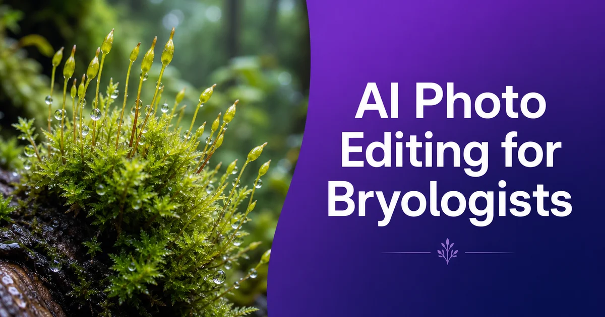
Bryology — the study of mosses, liverworts, and hornworts — relies on high-quality specimen photography at scales ranging from landscape habitat shots to individual cell structure for species identification, taxonomic description, ecological monitoring, and conservation assessment. With about 20,000 described moss species, 9,000 liverwort species, and 200 hornwort species worldwide, and new species still being described regularly from tropical and boreal regions, the ability to produce clear, detailed, standardized images across multiple magnification scales is fundamental to research progress. Every taxonomic description requires figure plates showing diagnostic features from the growth-form level down to leaf cell morphology. Revisionary work often demands re-imaging type specimens from herbaria spanning multiple continents.
The photographic challenges in bryology are distinctive and formidable. Most bryophytes are small — many common mosses have leaves less than two millimeters long. The cellular features that drive identification require compound microscope magnification. Specimens are often three-dimensional, with complex branching patterns, overlapping leaves, and sporophytes extending vertically from the gametophyte, making full-depth imaging impossible in a single exposure at any meaningful magnification. Color is often muted — greens, browns, and olive tones that are difficult to differentiate without careful lighting. And critically, many liverwort identification characters depend on oil bodies within leaf cells that degrade within hours to days of collection, requiring rapid photography of fresh material before these ephemeral features are lost.
AI photo editing tools directly address these challenges by automating post-processing steps that otherwise consume major research time. Background removal isolates specimens from the complex substrates they naturally colonize: bark, rock, soil, decaying wood, and often other bryophyte species growing in intimate mixture. Detail boost recovers the fine cellular and structural features that are diagnostically critical at high magnification. Batch processing standardizes images from multi-day field expeditions or herbarium visits where lighting equipment and conditions varied between sessions. For bryologists managing fieldwork, laboratory analysis, teaching responsibilities, and publication timelines at once, efficient image processing is not a luxury but a practical necessity for productive research.
- Background removal isolates bryophyte specimens from bark, rock, soil substrates, and the mixed-species growth communities they typically inhabit.
- AI enhancement sharpens diagnostically critical microscopic structures — leaf cell shape, costa anatomy, peristome ornamentation, and liverwort oil bodies — at the magnifications taxonomy requires.
- Magic Eraser removes mounting artifacts, soil contamination, and fungal overgrowth without altering the diagnostic morphological features of irreplaceable herbarium specimens.
- Batch processing standardizes images from field expeditions and herbarium visits spanning different institutions, equipment setups, and lighting conditions.
- Multi-scale figure plates from habitat context to cellular detail are exported at 300 DPI to meet journal publication requirements for taxonomic descriptions.
Multi-scale photography challenges and AI-assisted post-processing
Bryophyte documentation uniquely requires photography spanning at least four magnification scales: habitat context showing the species in its ecological setting, colony growth form showing branching pattern and shoot arrangement, individual leaf or thallus lobe morphology, and cellular detail at compound microscope magnification. A complete species record may require images at 1x, 10x, 40x, and 400x magnification, each demanding different equipment, lighting, and processing workflows. Focus stacking is key at the macro and stereomicroscope levels where three-dimensional moss cushions and erect sporophytes extend well beyond the available depth of field, generating dozens of source frames per view that must be aligned and merged.
AI post-processing integrates into each magnification level's workflow. At the macro level, background removal separates the target species from its growth companions. A critical function because bryophytes rarely grow in monospecific stands. At stereomicroscope magnification, focus-stacking artifacts (halos, edge fringing, and doubled edges from imperfect alignment) are cleanly eliminated alongside the background. At compound microscope magnification, AI boost sharpens cell wall outlines, papillae on cell surfaces, and the contents of individual cells that are critical for liverwort identification. Color normalization across all scales ensures that the green of the same species photographed through different optical systems appears consistent in the final figure plate.
Lighting for bryophyte photography requires particular care because the subjects are mostly green, often translucent, and frequently wet in their natural state. Moss leaves photographed with transmitted light reveal cell structure beautifully but lose their natural growth-form context. Reflected light preserves growth form but cannot show cellular detail. Field photography contends with forest-floor shade, canopy dappling, and the moisture that keeps bryophytes photosynthetically active but creates glare on wet surfaces. AI color correction and exposure normalization compensate for these variable conditions, producing consistent results from images taken under very different lighting settings.
- Documentation spans at least four magnification scales — habitat context, colony growth form, leaf morphology, and cellular detail — each requiring different equipment and processing.
- Focus stacking at macro and stereomicroscope levels generates dozens of frames per view; AI cleanly eliminates stacking artifacts and mixed-species backgrounds.
- Color normalization ensures the same species appears consistent across images taken through different optical systems and lighting environments.
- AI enhancement at compound microscope magnification sharpens cell wall outlines, surface papillae, and intracellular contents critical for liverwort identification.
Enhancing diagnostic features for bryophyte taxonomic identification
Moss taxonomy relies on a hierarchy of morphological features that images must clearly resolve for scientific value. At the growth-form level, branching pattern (pinnate, dendroid, julaceous, or irregular), shoot orientation (erect, procumbent, or pendent), and the presence and form of specialized structures like paraphyllia and pseudoparaphyllia are primary characters. At the leaf level, shape, margin serration, costa length and structure, and the differentiation of alar cells at the leaf base are all diagnostically critical. At the cellular level, cell shape (linear, hexagonal, quadrate, or rhomboidal), wall thickness, and the presence of papillae or mammillae on cell surfaces distinguish genera and species. AI boost that increases local contrast and sharpness at each scale makes these features clearly visible in photographs that would otherwise require microscope examination to interpret.
Liverwort identification depends on characters that are photographically even more demanding. The leafy liverworts (Jungermanniales) use leaf insertion angle, underleaf form, and cellular oil bodies as primary diagnostic characters. Oil bodies — organelles within leaf cells that contain terpenoid compounds — vary enormously between genera in number per cell, shape (spherical, ellipsoidal, grape-cluster, or segmented), color, and surface texture (smooth, granular, or papillose). These structures degrade within hours to days after collection, making rapid photography of fresh material key. AI boost maximizes the visibility of oil body traits in photographs that must be taken quickly before the structures deteriorate, extracting maximum diagnostic information from necessarily imperfect source images.
Sporophyte characters — capsule shape, peristome tooth structure and ornamentation, calyptra form, and seta texture — are key for many moss identifications and for all higher-level classification. Peristome teeth are among the most complex and beautiful microscopic structures in any plant group, with elaborate lattice-work ornamentation that must be clearly documented at high magnification. The teeth are often less than half a millimeter long and their ornamentation features are measured in micrometers. AI detail boost recovers the fine ornamentation patterns that standard light microscopy photographs often render as blurred or indistinct, including the delicate cross-bars, papillae, and trabeculae that distinguish peristome types across moss families.
- Growth-form, leaf-level, and cellular characters form a multi-scale diagnostic hierarchy that AI enhancement renders visible at each magnification level.
- Liverwort oil bodies — ephemeral organelles that degrade within hours — require rapid photography where AI maximizes diagnostic visibility from necessarily imperfect source images.
- Peristome tooth ornamentation at sub-millimeter scales demands AI enhancement to recover fine lattice-work, papillae, and trabeculae that distinguish moss family classification.
- AI sharpening of leaf margin serration, costa structure, and alar cell differentiation provides the precision needed for genus and species-level identification from photographs.
Processing herbarium specimens and managing preservation artifacts
The world's bryophyte research collections — estimated at over 70 million herbarium specimens in institutions globally — represent an irreplaceable record of diversity collected over more than three centuries. Most bryophyte herbarium specimens are air-dried and stored in paper packets, a keeping method that maintains morphological features well but introduces specific photographic challenges. Dried specimens are compressed, often losing their three-dimensional growth form. Colors shift from living greens to browns and olive tones. Leaves may curl, overlap, or become detached from stems during decades of storage. Rehydrating specimens in water restores some three-dimensionality but changes the optical properties of tissues and may damage fragile material.
AI processing tools help maximize the information extractable from herbarium material without physical manipulation that risks damaging irreplaceable specimens. Boost applied to photographs of dried specimens increases the visibility of features obscured by compression. Branching pattern, leaf orientation, and capsule arrangement can often be determined from enhanced images of well-prepared specimens that would require physical dissection to see clearly in the original material. Background removal eliminates mounting paper, adhesive residue, and packet edges from images, producing clean specimen photographs suitable for digital herbarium databases and online spotting resources.
For historically major specimens — types described by foundational bryologists like Hedwig, Bridel, Schwaegrichen, and Mitten in the eighteenth and nineteenth centuries — physical condition is often poor, and the specimens cannot be freely handled or dissected. High-resolution photography with AI boost provides a way to extract maximum morphological information from this material non-destructively. Enhanced images of type specimens are increasingly important for taxonomic stability, providing modern researchers with the visual records needed to correctly apply species names originally described from text-only or minimally illustrated publications. Several major bryophyte herbaria are now systematically photographing and enhancing their type collections to make this critical material accessible worldwide.
- Air-dried herbarium specimens lose three-dimensionality and natural color; AI enhancement increases visibility of diagnostic features obscured by compression and color shift.
- Background removal eliminates mounting paper, adhesive residue, and packet edges for clean specimen images suitable for digital herbarium databases.
- Non-destructive high-resolution photography with AI enhancement extracts maximum information from irreplaceable historical type specimens that cannot be freely handled.
- Systematic AI-enhanced photography of type collections is making foundational bryological material accessible worldwide for taxonomic verification and revisionary research.
Citizen science, conservation monitoring, and educational outreach
Bryophyte conservation increasingly depends on citizen science contributions for distribution data. The quality of photographic records directly determines whether expert verification is possible. Most bryophyte observations submitted to platforms like iNaturalist cannot be identified to species level from photographs alone because the diagnostic characters are microscopic. However, AI-enhanced photography that maximizes the visibility of macroscopic characters — growth form, branching pattern, leaf shape and orientation — can often narrow spotting to genus level, which is enough for many conservation monitoring purposes. Improving the photographic quality of citizen science contributions through accessible AI tools expands the effective monitoring network for bryophyte conservation without requiring every contributor to own a microscope.
Conservation assessment for bryophytes faces the challenge that many species are rare, narrowly endemic, or associated with threatened habitats like old-growth forests, peatlands, and montane cloud forests. Documenting these species photographically during rapid biodiversity surveys requires efficient image processing that can handle the volume of specimens photographed in a single field day. AI batch processing — applying consistent background removal, color correction, and boost across hundreds of images — makes it practical to produce standardized records from intensive field campaigns where time and conditions did not allow careful studio-quality photography for every specimen.
Educational outreach for bryology faces the fundamental challenge of scale: the features that make bryophytes biologically fascinating and ecologically important are largely invisible to the unaided eye. AI-enhanced macro and microscope photography transforms functional research images into strong visual content that reveals the intricate architecture of moss leaves, the jewel-like clarity of liverwort oil bodies, and the geometric precision of peristome teeth. These enhanced images serve museum exhibits, university teaching materials, natural history publications, and social media science communication, building appreciation for a plant group that most people walk past without noticing despite bryophytes being present in virtually every terrestrial ecosystem on earth.
- AI-enhanced macroscopic photography enables citizen science contributors to submit bryophyte observations identifiable to genus level without requiring microscope access.
- Batch processing standardizes documentation from intensive field campaigns where rapid biodiversity surveys preclude studio-quality photography for each specimen.
- Conservation monitoring for rare and endemic bryophyte species benefits from efficient AI processing that handles the volume of specimens from targeted survey work.
- Educational outreach uses enhanced imagery to reveal the invisible micro-architecture of bryophytes to audiences who overlook them despite their ecological ubiquity.
How to do it
- 1
Photograph bryophyte specimens with standardized macro and microscope setups
Set up a macro photography station with diffused lighting for habit shots showing the overall growth form of moss cushions, liverwort thalli, or hornwort rosettes. Include a millimeter scale bar in every frame. For taxonomically critical features, switch to a stereomicroscope or compound microscope with a camera adapter to photograph leaf cell structure, capsule morphology, peristome teeth, and oil body traits in liverworts. Use focus stacking for three-dimensional structures like moss sporophytes and capsules where depth of field at high magnification captures only a fraction of the specimen.
- 2
Remove distracting substrates and isolate specimens for publication
Use Background Eraser to isolate bryophyte specimens from bark, rock, soil. The tangle of other bryophyte species they often grow among. Mosses and liverworts almost never grow in isolation. A single log may host a dozen species intermixed, and separating the target species visually from its neighbors is key for clear records. Background removal is mainly valuable for herbarium specimens where mounting paper, label edges. Adhesive residue can interfere with the visibility of diagnostic features like branching pattern, leaf orientation, and capsule arrangement.
- 3
Enhance fine cellular and structural details critical for identification
Use AI Enhance to sharpen the microscopic structures that drive bryophyte spotting. Leaf cell shape and papillosity, costa (midrib) structure and length, alar cells at the leaf base, capsule wall anatomy, peristome tooth ornamentation in mosses, and the ephemeral oil bodies that are the primary spotting character for many liverwort genera. Many of these features are measured in micrometers and require maximum image clarity for species-level spotting in taxonomic keys.
- 4
Clean up preparation artifacts and substrate contamination
Use Magic Eraser to remove mounting debris, soil particles, fungal hyphae growing over the specimen. Other contaminants that obscure diagnostic morphological features. This is mainly valuable for herbarium specimens collected decades ago where specimens were mounted with methods that now interfere with high-resolution photography. Heavy glue adhesion, paper packets that compress three-dimensional structures, and labels placed over portions of the specimen that cannot be physically moved without risking damage to the irreplaceable material.
- 5
Prepare multi-scale figure plates from habit to cellular detail
Arrange standardized views spanning multiple magnification levels. Habitat context, colony growth form, individual shoot or thallus, leaf shape, and cellular detail — into publication-ready figure plates with consistent spacing and labeling. Export at 300 DPI minimum for journal submission. For spotting guides and citizen science platforms, prepare extra web-optimized versions with key diagnostic features annotated to bridge the gap between what non-specialists photograph in the field and the microscopic characters that drive formal spotting.
Sources
- Photographic Techniques for Bryophyte Identification and Documentation — British Bryological Society
- Best Practices for Herbarium Specimen Imaging in Cryptogamic Collections — Smithsonian National Museum of Natural History
- Focus Stacking and Image Processing for Microscopic Bryophyte Structures — The Bryologist — American Bryological and Lichenological Society