
Retouche photo IA pour perfusionnistes : Documenter les circuits et équipements — Magic Eraser
Retouche photo experte pour perfusionnistes et équipes de perfusion cardiovasculaire. Outils IA pour les registres de circuits, la photographie d'équipement, la dé-identification conforme HIPAA et la création de matériel de formation.
SEO & Growth
Vérifié par Magic Eraser Editorial ·
Sommaire
- Dé-identification conforme HIPAA de la photographie de perfusion en salle d'opération
- Améliorer la photographie de circuits de dérivation pour la formation et la documentation de cas
- Simplification de circuits et isolation de composants ciblés pour les matériels éducatifs
- Présentations de conférences, soumissions de journaux et développement de portfolio professionnel
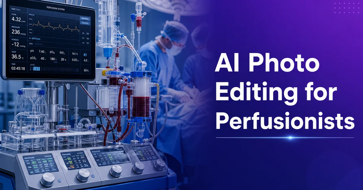
Les perfusionnistes cardiovasculaires manœuvrent les machines cœur-poumon qui maintiennent les patients en vie pendant la chirurgie à cœur ouvert et d'autres interventions nécessitant une circulation extracorporelle. La documentation de leur travail comporte des défis photographiques hautement spécialisés que les outils génériques de retouche photo n'ont jamais été conçus pour relever. Chaque cas de circulation extracorporelle produit une configuration de circuit qui constitue véritablement un système de maintien des fonctions vitales fait sur mesure. Un oxygénateur, un réservoir veineux, un filtre artériel, un échangeur thermique, des têtes de pompe et des mètres de tubulures interconnectées assemblés selon les besoins précis du patient et de l'intervention. Documenter ces configurations par la photographie est essentiel pour l'assurance qualité, la formation continue, la revue de cas, la formation des nouveaux perfusionnistes. Et pour contribuer à la littérature spécialisée qui fait progresser la pratique de la perfusion.
Le contexte photographique du travail de perfusion compte parmi les plus difficiles de toutes les professions. Les blocs opératoires sont éclairés par d'intenses scialytiques qui créent des ombres dures et des hautes lumières brûlées sur les surfaces réfléchissantes de la machine cœur-poumon. Le circuit de circulation extracorporelle lui-même est un assemblage dense de composants plastiques transparents et translucides reliés par des dizaines de lignes de tubulure presque impossibles à suivre visuellement sur une photographie. Les équipements de surveillance du patient, les appareils d'anesthésie, les instruments chirurgicaux et le personnel encombrent le champ de vision. Plus crucial encore, le bloc opératoire contient une abondance de renseignements médicaux protégés. Noms de patients sur les moniteurs, numéros de dossier médical sur les bracelets, visages du personnel et identifiants institutionnels — ce qui fait de chaque photographie un risque potentiel de violation de la HIPAA si elle est partagée sans dépersonnalisation rigoureuse.
Les outils de retouche photo par IA résolvent les défis spécifiques qui rendent la photographie de perfusion difficile et sensible sur le plan de la conformité. La suppression d'arrière-plan et l'effacement d'objets permettent une dépersonnalisation HIPAA rigoureuse en éliminant des images les éléments identifiant le patient et les détails propres à l'établissement avant tout usage éducatif ou de publication. L'amélioration par IA restaure les détails du circuit. Étiquettes des composants, raccords de tubulure, niveaux et couleurs des fluides — que l'éclairage du bloc opératoire fait disparaître ou masque. La suppression sélective d'objets simplifie les photographies de circuits complexes en isolant le composant ou le raccord précis à documenter de l'enchevêtrement environnant de lignes et d'équipements. Ce guide couvre le flux de travail complet des perfusionnistes, depuis la prise de vue au bloc opératoire jusqu'à un rendu dépersonnalisé, amélioré et prêt à publier.
- Background Eraser supprime les environnements de bloc opératoire contenant moniteurs de patients, personnel et identifiants d'établissement, et les remplace par des arrière-plans cliniques nets pour une documentation conforme à la HIPAA.
- AI Enhance restaure le texte des étiquettes de composants, les détails des raccords de tubulure et les dégradés de couleur des fluides que l'éclairage du bloc opératoire efface sur les images de téléphone.
- Magic Eraser supprime sélectivement les tubulures superflues et les équipements pour isoler des composants précis du circuit, rendant la documentation de formation plus claire et plus ciblée.
- La photographie de configuration de circuit avec des arrière-plans cohérents crée des archives de documentation de cas professionnelles, adaptées à la revue d'assurance qualité et aux dossiers de l'établissement.
- L'export par lots produit des versions prêtes pour la présentation, de qualité de publication et en résolution d'archivage à partir d'un seul master édité, pour les conférences, les revues et les dossiers de cas.
Dé-identification conforme HIPAA de la photographie de perfusion en salle d'opération
L'étape la plus critique de la retouche photographique de perfusion est une dépersonnalisation rigoureuse qui supprime les dix-huit catégories de renseignements médicaux protégés définies par la règle de confidentialité de la HIPAA avant toute utilisation d'une image hors de l'équipe clinique immédiate. Les photographies de bloc opératoire captent couramment des noms de patients affichés sur les écrans de surveillance et les dossiers d'anesthésie, des numéros de dossier médical sur les bracelets et étiquettes, des dates d'intervention visibles à l'arrière-plan, des traits du visage de patients sédatés partiellement visibles au-delà des champs chirurgicaux. Ainsi que des identifiants institutionnels sur les étiquettes d'équipement et les badges du personnel. Un seul identifiant négligé dans une présentation de congrès ou un cas clinique publié peut constituer une violation de la HIPAA aux conséquences juridiques et professionnelles majeures pour le perfusionniste et son établissement.
Background Eraser offre l'approche la plus complète de la dépersonnalisation en supprimant tout le décor du bloc opératoire et en le remplaçant par un arrière-plan clinique neutre qui conserve le contexte médical spécialisé sans aucun détail identifiant. Cette approche est plus fiable que le floutage ou le caviardage sélectifs car elle élimine le risque de manquer un petit identifiant dans une scène complexe. Si tout l'arrière-plan est remplacé, chaque élément d'arrière-plan est supprimé, qu'il ait ou non été identifié individuellement comme sensible. L'arrière-plan de remplacement doit évoquer un cadre clinique. Des tons gris neutres ou blanc-bleuté avec de subtiles silhouettes d'équipement médical — afin que la photographie du circuit conserve son contexte spécialisé sans le risque de conformité du véritable bloc opératoire.
Pour les photographies où il importe de conserver une partie du contexte du bloc opératoire — comme les images montrant la relation spatiale entre le circuit de circulation extracorporelle et le champ chirurgical — Magic Eraser permet une suppression ciblée d'identifiants précis tout en préservant la composition globale de la scène. Cette approche sélective exige un examen attentif de chaque élément de l'image : les écrans des moniteurs doivent être effacés ou remplacés par des affichages génériques, tout texte visible sur les dossiers du patient doit être supprimé, les visages du personnel doivent être masqués. Les logos institutionnels ou noms de service sur les murs et les équipements doivent être éliminés. Un processus de révision en deux passes — effacement initial suivi d'un balayage systématique à l'aide d'une liste de contrôle des identifiants HIPAA — garantit une dépersonnalisation complète avant que l'image ne quitte le cadre clinique.
- Background Eraser supprime des environnements entiers de bloc opératoire pour une fiabilité maximale de dépersonnalisation, en éliminant les dix-huit catégories d'identifiants HIPAA en une seule opération.
- Les arrière-plans cliniques neutres de remplacement conservent un contexte médical professionnel sans aucun identifiant de patient, de personnel ou d'établissement issu du cadre d'origine.
- Magic Eraser offre une suppression ciblée lorsque le contexte du bloc opératoire doit être partiellement préservé, exigeant une révision systématique au regard des listes de contrôle des identifiants HIPAA.
- La dépersonnalisation en deux passes — effacement initial et balayage systématique par liste de contrôle — garantit une conformité complète avant tout usage éducatif ou de publication.
Améliorer la photographie de circuits de dérivation pour la formation et la documentation de cas
La photographie de circuits de circulation extracorporelle présente des défis d'amélioration uniques car l'information visuelle la plus importante se trouve dans des composants transparents et translucides que les téléphones gèrent mal. L'oxygénateur — le composant qui assure les échanges gazeux et remplace la fonction des poumons du patient — est logé dans une coque plastique transparente à travers laquelle le faisceau de fibres de la membrane et les motifs d'écoulement du sang sont partiellement visibles. Le réservoir veineux recueille le sang revenant du patient à travers un boîtier transparent qui permet au perfusionniste de surveiller le volume sanguin et de repérer les bulles d'air. Les filtres de ligne artérielle ont des boîtiers transparents qui révèlent la maille du filtre et toute particule capturée. Dans tous ces composants, l'information visuelle critique se trouve derrière ou à l'intérieur du plastique transparent. Les téléphones peinent à résoudre ce détail sous l'éclairage mixte du bloc opératoire.
AI Enhance restaure le détail de ces composants transparents en améliorant le contraste et la netteté à l'intérieur des boîtiers plastiques tout en gérant les reflets spéculaires qui créent des zones surexposées sur les surfaces plastiques courbes. Après amélioration, les photographies de l'oxygénateur montrent la disposition des fibres de la membrane et les motifs de distribution du sang qui indiquent un bon fonctionnement. Les images du réservoir révèlent des niveaux de fluide précis et l'absence ou la présence de bulles d'air avec assez de netteté pour un usage de formation. Les photographies du filtre montrent la capture de particules pouvant faire partie d'une revue qualité post-intervention. L'amélioration accroît aussi la lisibilité des étiquettes de composants. Nom du fabricant, numéro de modèle, numéro de lot et débit maximal imprimés sur les boîtiers plastiques — essentiels pour les dossiers de cas et les signalements d'incident si un problème de performance d'un composant est identifié ultérieurement.
La photographie des raccords de tubulure est un autre domaine où l'amélioration accroît grandement la qualité de la documentation. Les points de raccordement entre composants du circuit sont l'endroit où les erreurs d'assemblage surviennent le plus souvent. Les supports de formation doivent montrer clairement l'orientation correcte des connecteurs, les mécanismes de verrouillage et la confirmation visuelle d'une fixation sûre. Sur les photographies de bloc opératoire, ces raccords sont souvent masqués par des ombres, des tubulures concurrentes. Et par la confusion visuelle de plusieurs connecteurs d'aspect semblable très proches. L'amélioration affine le détail des connecteurs, améliore la récupération des ombres pour que la géométrie du raccord soit visible même dans les zones mal éclairées, et fait ressortir le code couleur des segments de tubulure qui identifie les lignes artérielles, les lignes veineuses, les circuits de cardioplégie et les lignes de purge. Des distinctions de couleur essentielles à un fonctionnement sûr du circuit mais souvent effacées sur les photos de téléphone.
- L'amélioration des composants transparents révèle les motifs de la membrane de l'oxygénateur, les niveaux de fluide du réservoir et le détail de capture du filtre à travers des boîtiers plastiques que les téléphones ne peuvent résoudre.
- La lisibilité des étiquettes de composants s'améliore pour les spécifications du fabricant, les numéros de lot et les débits, essentiels à la documentation des cas et au signalement d'incidents.
- La photographie des raccords de tubulure montre l'orientation correcte des connecteurs et les mécanismes de verrouillage assez clairement pour les supports de formation sur la sécurité d'assemblage du circuit.
- L'amélioration du code couleur restaure les distinctions des lignes artérielle, veineuse, de cardioplégie et de purge, essentielles à un fonctionnement sûr mais effacées sous l'éclairage du bloc.
Simplification de circuits et isolation de composants ciblés pour les matériels éducatifs
Un circuit de circulation extracorporelle entièrement assemblé est l'un des systèmes les plus complexes visuellement en médecine, avec des dizaines de raccords de tubulure créant un réseau tridimensionnel que même des perfusionnistes expérimentés peinent à suivre sur une photographie en deux dimensions. Pour les supports de formation qui doivent expliquer des composants individuels ou des sous-systèmes précis du circuit, cette complexité est un obstacle à l'apprentissage plutôt qu'une aide. Les nouveaux étudiants en perfusion, devant la photographie d'un circuit complet, sont submergés par la densité d'information visuelle et ne peuvent identifier l'élément précis abordé. Magic Eraser y remédie en supprimant les tubulures non pertinentes, les lignes de surveillance. Et les équipements afin d'isoler le composant ou le sous-système enseigné, réduisant la charge cognitive et concentrant l'attention là où l'instruction est dirigée.
L'isolement des sous-systèmes est particulièrement utile pour enseigner les circuits fonctionnels distincts d'un montage de circulation extracorporelle. La boucle artério-veineuse principale, le circuit de délivrance de la cardioplégie, le système d'ultrafiltration. La ligne de purge ventriculaire sont des sous-systèmes fonctionnels distincts qui se trouvent physiquement entremêlés sur la machine cœur-poumon. En supprimant sélectivement les tubulures et composants appartenant aux autres sous-systèmes, un perfusionniste formateur peut créer des photographies ne montrant par exemple que le circuit de cardioplégie, avec sa tête de pompe dédiée, son échangeur thermique, sa ligne de délivrance. Et ses points de surveillance visibles sur un arrière-plan net. Cette approche d'isolement des sous-systèmes transforme des photographies complexes de circuit entier en une bibliothèque d'images d'enseignement ciblées, chacune mettant en valeur un élément fonctionnel avec la clarté qu'exige un enseignement efficace.
Les présentations en construction séquentielle exploitent cette capacité d'isolement pour enseigner l'assemblage du circuit en montrant le système gagner en complexité un composant à la fois. En partant d'une photographie du seul oxygénateur, chaque image suivante de la série ajoute un composant de plus. Réservoir veineux, filtre artériel, têtes de pompe, raccords de surveillance — jusqu'à ce que l'image finale montre le circuit complet avec tous les éléments visibles. Cette approche additive, créée en partant d'un circuit entièrement photographié puis en retirant progressivement moins d'éléments à chaque version, aide les étudiants à comprendre non seulement ce que sont les composants mais comment ils se relient entre eux dans l'espace et la fonction. Le résultat est une ressource de formation qui construit la compréhension progressivement plutôt que de présenter d'un coup toute la complexité d'un circuit de circulation extracorporelle.
- Magic Eraser supprime les tubulures non pertinentes et les équipements pour isoler des composants précis, réduisant la charge cognitive des étudiants en perfusion qui apprennent les éléments individuels du circuit.
- L'isolement des sous-systèmes sépare les circuits artério-veineux, de cardioplégie, d'ultrafiltration et de purge en images d'enseignement individuelles à partir d'une seule photographie de circuit entier.
- Les présentations en construction séquentielle montrent l'assemblage du circuit un composant à la fois, créées en retirant progressivement moins d'éléments d'un circuit entièrement photographié.
- Les photographies de composants ciblées transforment des images de bloc opératoire visuellement complexes en supports pédagogiques clairs et lisibles, adaptés aux diapositives de cours et aux laboratoires de simulation.
Présentations de conférences, soumissions de journaux et développement de portfolio professionnel
Les présentations de perfusion à l'AmSECT, à l'ELSO et aux congrès régionaux s'appuient de plus en plus sur des preuves photographiques pour étayer les cas cliniques, les démonstrations de technique et les démarches d'amélioration de la qualité, faisant de la qualité d'image un facteur de l'accueil réservé au travail d'un perfusionniste par ses pairs spécialisés. Des photographies aux arrière-plans nets, à la visibilité des composants améliorée. Et à l'étiquetage clair projettent professionnalisme et compétence technique, tandis que des clichés mal éclairés de bloc opératoire aux arrière-plans encombrés et aux étiquettes illisibles minent la crédibilité d'un travail clinique par ailleurs excellent. La retouche par IA comble l'écart entre les photographies que les perfusionnistes peuvent réalistement capturer sous la pression temporelle des cas cliniques et la qualité d'image attendue dans les présentations spécialisées.
Les soumissions aux revues et publications ont des exigences d'image précises que la retouche par IA aide les perfusionnistes à satisfaire de façon constante. Le Journal of ExtraCorporeal Technology, Perfusion et d'autres publications spécialisées exigent des images haute résolution avec une dépersonnalisation complète du patient, une annotation claire des composants et une qualité spécialisée comparable aux images de la littérature existante. De nombreux perfusionnistes ont de précieuses expériences de cas et des configurations de circuit inédites dignes de publication mais manquent des ressources photographiques pour produire des images de qualité de publication à partir de leurs dossiers de bloc opératoire. L'amélioration par IA transforme les photos de téléphone en images conformes aux normes éditoriales. Une dépersonnalisation systématique assure la conformité HIPAA pour le processus d'examen par le CER qui régit la publication des cas cliniques. Pouvoir produire des images prêtes à publier à partir de dossiers de cas courants abaisse la barrière à la contribution à la littérature spécialisée.
Le développement de portfolios professionnels est une application émergente où les perfusionnistes documentent leur expérience clinique, leurs compétences spécialisées. Et leur familiarité avec les équipements à des fins d'avancement de carrière, notamment candidatures, examens d'accréditation et certification de spécialiste. Un portfolio montrant des configurations de circuit nettes et bien documentées pour une variété de types d'intervention. Pontage coronarien, remplacement valvulaire, chirurgie aortique, cas pédiatriques, mise en place d'ECMO — témoigne de l'étendue de l'expérience bien mieux qu'un curriculum vitae ne listant que des nombres de cas. La retouche par IA garantit que les images du portfolio sont toujours professionnelles, soigneusement dépersonnalisées et clairement documentées avec le type de cas et la configuration du circuit. Cette documentation visuelle de la compétence clinique est particulièrement précieuse pour les perfusionnistes cherchant un poste dans des centres spécialisés où l'expérience d'un équipement précis et le volume d'interventions sont des critères d'embauche.
- Les présentations en congrès avec des photographies de circuit nettes et améliorées projettent un professionnalisme et une crédibilité que les clichés de bloc opératoire aux arrière-plans encombrés ne peuvent égaler.
- Des images de qualité de publication issues de la documentation courante des cas abaissent la barrière pour les perfusionnistes souhaitant soumettre des cas cliniques et des configurations inédites aux revues spécialisées.
- Les portfolios professionnels montrant des configurations de circuit variées et bien documentées démontrent l'étendue clinique et l'expérience des équipements pour l'avancement de carrière et l'accréditation.
- Des normes de retouche par IA cohérentes sur l'ensemble de la documentation des cas créent des bibliothèques d'images institutionnelles précieuses pour les revues d'assurance qualité et l'accréditation des programmes de formation.
How to do it
- 1
Photograph cardiopulmonary bypass circuits with clear component identification
Set up your circuit photography to capture the complete heart-lung machine configuration with each component clearly visible and identifiable. Position the camera at a slight angle that shows the oxygenator, venous reservoir, arterial filter, heat exchanger. Pump heads in spatial relationship to each other, with tubing connections traceable between components. Use even, diffused lighting that minimizes reflections on the transparent plastic components while maintaining visibility of fluid levels and color changes within the circuit. Label photographs right away with patient-de-identified case information including circuit type, oxygenator model, and any non-standard configurations used.
- 2
Remove patient-identifiable information and operating room backgrounds
Use Background Eraser and Magic Eraser to remove all elements that could identify patients, staff, or specific facility locations from circuit and equipment photographs. Operating room settings contain monitors displaying patient vitals, name bands visible on draped arms, staff ID badges. Facility-specific equipment labels that constitute protected health information under HIPAA. Replace operating room backgrounds with clean clinical settings that maintain the expert medical context without any identifying details. This de-spotting step is mandatory before any educational, conference, or publication use of perfusion photography.
- 3
Enhance circuit detail for tubing connections, fluid levels, and component labels
Apply AI Enhance to improve the visibility of critical circuit details that are difficult to capture clearly with phone cameras in operating room lighting conditions. Transparent and translucent circuit components — oxygenator membranes, venous reservoirs, arterial line filters — are mainly challenging because the camera struggles to resolve detail through multiple layers of plastic housing. Boost recovers the text on component labels, makes tubing connection points clearly visible for training records. Improves the visibility of fluid color gradients that indicate oxygenation status. These enhanced details transform quick operating room snapshots into records-quality images suitable for educational displays and quality assurance records.
- 4
Clean up tubing clutter and isolate specific circuit components for focused documentation
Use Magic Eraser to remove extraneous tubing runs, cable bundles. Equipment that obscure the specific circuit component or connection being documented. A cardiopulmonary bypass circuit involves dozens of tubing connections, monitoring lines. Drug infusion sets that create visual complexity making it difficult to focus attention on the particular element being documented. Selectively removing non-relevant tubing and equipment from the photograph allows viewers to follow the specific fluid path or connection point being highlighted without the cognitive burden of tracing lines through a tangle of similar-looking tubes.
- 5
Export perfusion images for case documentation, training presentations, and publications
Save edited perfusion photographs at full resolution for institutional case records archives, then create display-ready versions for educational lectures, simulation training materials, and conference submissions. Training displays benefit from consistent backgrounds and enhanced component visibility that make circuit configurations readable on projected screens in lecture halls. Publication submissions require high-resolution images with complete de-spotting records confirming HIPAA compliance. Batch export handles the multiple images from a single case. Circuit setup, mid-case configurations, and post-case records — maintaining consistent visual quality across the image series.
Sources
- Standards of Practice for Perfusion: AmSECT Position Statements — American Society of ExtraCorporeal Technology
- Medical Photography Standards and Patient Privacy Requirements — U.S. Department of Health and Human Services
- Visual Documentation in Clinical Education and Simulation Training — Society for Simulation in Healthcare