
Retouche photo IA pour myrmécologistes — Magic Eraser
Comment les myrmécologistes utilisent la retouche photo IA pour les registres de spécimens de fourmis, la photographie taxonomique et les publications de recherche. Supprimez les artefacts d'épingles, améliorez les détails morphologiques et créez des planches prêtes à publier pour la systématique des fourmis.

SEO & Growth
Vérifié par Magic Eraser Editorial ·
Sommaire
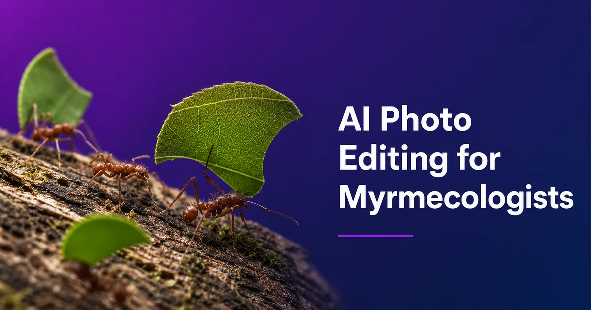
La myrmécologie — l'étude scientifique des fourmis — repose sur une photographie de spécimens de haute qualité couvrant des échelles allant des relevés d'habitat sur le terrain aux structures corporelles individuelles mesurées en centièmes de millimètre. Avec plus de 16 000 espèces de fourmis décrites et entre 6 000 et 8 000 espèces estimées encore en attente de description formelle, la demande d'imagerie de spécimens claire et standardisée n'a jamais été aussi forte. Chaque description d'espèce nouvelle requiert des planches de figures détaillées montrant les caractères morphologiques diagnostiques selon plusieurs vues standardisées, et la révolution des bases de données de taxonomie numérique. Principalement AntWeb, qui héberge désormais des images de plus de 90 000 spécimens de fourmis — a rendu les enregistrements photographiques aussi fondamentaux pour la systématique des fourmis que les spécimens physiques eux-mêmes.
Les défis photographiques en myrmécologie découlent de l'échelle et des besoins de standardisation. De la simple diversité du matériel qui doit être photographié. La plupart des ouvrières mesurent entre deux et quinze millimètres de long, ce qui les place dans la gamme techniquement exigeante où la macrophotographie à fort grossissement est nécessaire mais où les spécimens sont assez grands pour avoir une forme tridimensionnelle complexe. Une seule ouvrière peut présenter des motifs de sculpture sur la surface de la tête, des dents le long de la marge des mandibules, des épines projetées depuis le propodéum, et des soies (setae) ressemblant à des poils couvrant le corps. Tout cela doit être clairement résolu et distingué sur des photographies qui seront examinées à un zoom extrême par des taxonomistes du monde entier. L'empilement de mises au point (focus stacking) est essentiel mais introduit des artefacts. L'éclairage doit être assez diffus pour révéler la sculpture de surface sans aplatir la forme tridimensionnelle.
Les outils d'édition photo par IA répondent à ces défis en automatisant des étapes de post-traitement laborieuses qui consomment autrement une part substantielle du temps de recherche myrmécologique. La suppression de l'épingle et du montage produit des images de spécimens nettes adaptées aux bases de données et aux publications. L'amélioration des détails restitue les structures morphologiques fines — denture mandibulaire, motifs de sculpture, traits de pilosité — qui déterminent l'identification des espèces. La normalisation des couleurs compense la variation spectaculaire de l'apparence des spécimens causée par les différentes méthodes de conservation, les âges et les conditions photographiques. Pour les myrmécologues gérant à la fois des collections de terrain, des visites de musée, des charges d'enseignement. Et des échéances de publication, un traitement d'image efficace assisté par IA accélère directement le rythme de la recherche taxonomique.
- La suppression de l'épingle, du point-montage et de l'arrière-plan produit des images de spécimens nettes répondant aux protocoles d'imagerie standardisés d'AntWeb et d'autres bases de données de taxonomie numérique.
- L'amélioration par IA affine les structures morphologiques diagnostiques essentielles. Denture mandibulaire, épines propodéales, forme du pétiole, sculpture et pilosité — aux niveaux de grossissement requis par la taxonomie.
- La normalisation des couleurs compense les artefacts de conservation, l'assombrissement lié à l'âge et l'éclairage variable entre des spécimens photographiés dans différentes institutions et à différentes époques.
- Le nettoyage des artefacts d'empilement de mises au point élimine les halos, le dédoublement des bords et les franges qui compromettent la netteté des images composites à fort grossissement.
- Les planches de figures multi-vues en orientations dorsale, latérale et de face sont exportées à 300 DPI pour la soumission aux revues et la contribution aux bases de données taxonomiques.
Imagerie standardisée multi-vues et post-traitement assisté par IA
La photographie taxonomique des fourmis suit des protocoles de standardisation stricts développés sur des décennies afin que les images de différents chercheurs, institutions et équipements puissent être comparées de manière pertinente. Les trois vues requises — dorsale (regard plongeant sur le spécimen directement par-dessus), latérale (vue de profil depuis le côté, à la ligne médiane du spécimen). De face (tête vue directement de l'avant) — révèlent chacune des caractères diagnostiques différents. La vue dorsale montre la forme de la tête, le contour du mésosome et les proportions relatives des segments corporels. La vue latérale révèle les épines propodéales, les profils du pétiole et du postpétiole, et l'architecture corporelle globale. La vue de face expose la forme des mandibules, la structure du clypéus, la position des yeux et les points d'insertion antennaire. Les trois vues doivent être capturées avec le spécimen dans une orientation précisément contrôlée. Cela requiert un équipement de mise en scène spécialisé et beaucoup de patience.
Le post-traitement par IA s'intègre au flux de travail de chaque vue en plusieurs points. Après que l'empilement de mises au point a fusionné les 30 à 80 images capturées par vue en un seul composite entièrement net, l'IA supprime les artefacts d'empilement qui apparaissent invariablement. Halos autour des bords à fort contraste, bords dédoublés dus à de légères erreurs d'alignement entre images, et franges autour de structures saillantes comme les épines et les antennes qui bougent légèrement entre les expositions sur les spécimens anciens aux articulations relâchées. La suppression de l'épingle est l'opération de nettoyage la plus universelle : chaque spécimen épinglé montre l'épingle en acier inoxydable traversant le côté droit du mésosome, souvent avec de l'adhésif visible. Elle doit être supprimée pour des images de qualité publication tout en conservant la surface du mésosome qu'elle traverse.
La standardisation de l'arrière-plan est essentielle pour la contribution aux bases de données. AntWeb et les plateformes similaires affichent les images de spécimens sur des arrière-plans blancs unis avec une qualité d'éclairage constante. Les images soumises avec des arrière-plans encombrés ou incohérents sont soit rejetées, soit nécessitent une retouche manuelle chronophage. L'IA supprime non seulement l'arrière-plan physique du dispositif photographique, mais aussi le substrat de montage. Plateformes de mise en scène d'épingles, triangles de papier des points-montages et coton utilisés dans certaines préparations muséales — produisant des images de spécimens isolées qui répondent aux normes des bases de données sans les heures de masquage manuel que ces formes complexes et irrégulières exigeraient autrement.
- Trois vues standardisées — dorsale, latérale et de face — révèlent chacune des caractères diagnostiques différents essentiels à l'identification au niveau de l'espèce en taxonomie des fourmis.
- L'empilement de 30 à 80 images par vue introduit des artefacts que l'IA élimine proprement — halos, dédoublement des bords et franges autour des épines et antennes saillantes.
- La suppression de l'épingle à travers le mésosome est la tâche de nettoyage la plus universelle, préservant la surface corporelle autour du point d'insertion tout en effaçant la tige d'acier et l'adhésif.
- La standardisation de l'arrière-plan en blanc uni répond aux protocoles de soumission d'AntWeb et des bases de données qui exigent des normes d'imagerie cohérentes pour tous les spécimens contribués.
Améliorer les caractères morphologiques diagnostiques pour l'identification des fourmis
L'identification des espèces de fourmis repose sur une hiérarchie précise de caractères morphologiques que les photographies doivent résoudre clairement pour avoir une utilité scientifique. Au niveau du genre, la forme des mandibules. Le nombre, la taille et la disposition des dents le long de la marge masticatrice — est souvent le caractère diagnostique principal, certains genres étant définis par deux dents et d'autres par douze ou plus. La structure du clypéus, la forme des carènes frontales et le nombre d'articles antennaires distinguent davantage les genres. Au niveau de l'espèce, des caractères plus fins prévalent : la forme et la longueur spécifiques des épines propodéales, le profil des nœuds du pétiole et du postpétiole, la densité et le motif de la sculpture de surface (rugueuse, ponctuée, réticulée ou lisse). Le type, la densité et la distribution de la pilosité corporelle (pilosité et pubescence). L'amélioration par IA augmente le contraste local et la netteté pour rendre chacun de ces systèmes de caractères clairement lisible sur les photographies.
La sculpture de surface est surtout difficile à photographier parce qu'elle existe sous forme de texture tridimensionnelle très peu profonde sur la surface du corps. Les rugae — les crêtes saillantes qui couvrent la tête et le mésosome de nombreux genres de fourmis — peuvent ne s'élever que de quelques micromètres au-dessus de la surface environnante. Leur visibilité dépend entièrement de l'angle et de la qualité de l'éclairage par rapport à la surface. Les ponctuations — de minuscules dépressions circulaires dispersées sur la cuticule — sont encore plus subtiles. L'amélioration par IA compense l'éclairage imparfait en augmentant le micro-contraste qui rend ces caractères de surface visibles, restituant le détail de la sculpture dans les zones où l'angle d'éclairage d'origine se trouvait être parallèle à la surface et rendait la texture invisible.
La pilosité — le système des poils corporels — est l'un des systèmes de caractères les plus importants sur le plan diagnostique et les plus difficiles à photographier en taxonomie des fourmis. Les fourmis portent plusieurs types de poils corporels : des soies dressées (setae) qui se projettent perpendiculairement à la surface du corps, une pubescence couchée ou appliquée qui repose à plat contre la cuticule. Des formes de poils spécialisées sur des régions corporelles spécifiques. La distinction entre ces types de poils, leur densité, leur motif de distribution. Leur forme précise (droite, courbée, ramifiée ou claviforme) sépare des espèces étroitement apparentées. Ces structures sont souvent translucides et très fines. Une seule soie peut ne mesurer que quelques micromètres de diamètre — ce qui les rend difficiles à photographier clairement et quasi invisibles devant tout sauf un arrière-plan parfaitement net et contrasté.
- Le nombre et la disposition des dents mandibulaires est souvent le caractère diagnostique principal au niveau du genre — l'amélioration par IA doit résoudre les dents individuelles le long de la marge masticatrice.
- La sculpture de surface ne s'élève que de quelques micromètres au-dessus de la cuticule ; l'amélioration du micro-contraste par IA restitue le détail là où l'angle d'éclairage d'origine rendait la texture invisible.
- La forme et la longueur des épines propodéales, les profils du pétiole et la forme du postpétiole sont des caractères au niveau de l'espèce qui exigent une définition nette des bords sur les photographies en vue latérale.
- Les types de pilosité — soies dressées, pubescence appliquée et poils régionaux spécialisés — sont translucides et d'une épaisseur micrométrique, exigeant des arrière-plans nets et une netteté d'image maximale.
Traitement des collections muséales et gestion des artefacts de conservation
Les collections de fourmis du monde — estimées à plus de 100 millions de spécimens dans les musées d'histoire naturelle et les institutions de recherche à l'échelle mondiale — représentent une archive irremplaçable de la biodiversité accumulée sur plus de 250 ans d'efforts de collecte. Ces spécimens sont la preuve physique sous-jacente à chaque nom d'espèce publié, et les spécimens types. Les individus spécifiques qui servent de standard de référence pour chaque nom d'espèce — sont les objets scientifiquement les plus précieux de toute collection. Photographier les spécimens types pour les bases de données numériques est devenu une priorité pour les grands musées, mais l'état du matériel historique présente des défis photographiques constants : adhésifs anciens, épingles corrodées, étiquettes décolorées masquant des parties du corps. La détérioration physique de spécimens qui peuvent avoir plus d'un siècle.
Les outils de traitement par IA maximisent l'information extractible du matériel muséal sans manipulation physique risquant d'endommager des spécimens irremplaçables. L'amélioration appliquée aux photographies de spécimens anciens accroît la visibilité des caractères de surface masqués par les changements liés à l'âge. Assombrissement cuticulaire, accumulation de poussière et de résidus d'adhésif, et perte générale de contraste de surface qui se produit lorsque les spécimens s'oxydent au fil des décennies. La suppression de l'épingle est surtout précieuse pour les spécimens historiques où l'épingle est corrodée, tordue, ou a déposé une coloration de vert-de-gris sur la surface du spécimen. L'IA supprime l'artefact de l'épingle tout en conservant, voire en améliorant, la visibilité des caractères morphologiques immédiatement autour du point d'insertion de l'épingle.
Les campagnes de photographie de spécimens dans les grands musées peuvent impliquer l'imagerie de milliers de spécimens lors d'une seule visite de recherche, sous pression temporelle avec l'équipement d'éclairage disponible dans l'institution hôte. Le traitement par lots de l'IA standardise les résultats de ces séances d'imagerie intensives, appliquant une suppression d'arrière-plan et une correction des couleurs cohérentes. Et une amélioration sur des centaines d'images capturées dans des conditions variables. Cette capacité de traitement par lots transforme l'économie de la numérisation des fonds muséaux. Un chercheur en visite peut se concentrer sur la capture d'une couverture maximale plutôt que de passer du temps à perfectionner les images individuelles sur place, confiant que le post-traitement par IA portera l'ensemble du jeu au standard de publication.
- Plus de 100 millions de spécimens de fourmis dans les collections mondiales fournissent la preuve physique de chaque nom d'espèce publié — les spécimens types sont scientifiquement irremplaçables.
- L'amélioration par IA restitue les caractères de surface masqués par l'assombrissement lié à l'âge, les résidus d'adhésif, l'accumulation de poussière et la perte générale de contraste de la cuticule oxydée.
- Les épingles corrodées ou tachées de vert-de-gris sur les spécimens historiques sont supprimées tout en préservant le détail morphologique autour du point d'insertion.
- Le traitement par lots standardise l'imagerie issue de visites muséales intensives où des milliers de spécimens sont capturés dans des conditions variables et sous pression temporelle.
Relevés écologiques, science citoyenne et sensibilisation éducative
L'écologie des fourmis s'appuie de plus en plus sur l'identification photographique pour les relevés de biodiversité à grande échelle où collecter chaque spécimen est impraticable, indésirable ou interdit. Pièges photographiques, macrophotographie au smartphone et protocoles standardisés d'échantillonnage photo génèrent des milliers d'images par étude qui doivent être triées et identifiées. Et tandis que l'identification automatisée des espèces basée sur l'IA progresse rapidement, la qualité des images d'entrée limite fondamentalement la précision de l'identification. L'amélioration photo par IA appliquée aux images collectées sur le terrain avant qu'elles n'entrent dans les pipelines d'identification améliore la résolution des caractères diagnostiques, augmentant la proportion d'images identifiables au niveau de l'espèce plutôt que de s'arrêter au genre ou à la sous-famille. Pour les études écologiques suivant la composition des communautés, la différence entre une identification au niveau de l'espèce et au niveau du genre peut déterminer si des conclusions écologiques pertinentes sont possibles.
Les plateformes de science citoyenne comme iNaturalist reçoivent des centaines de milliers d'observations de fourmis chaque année. Le taux d'identification des photos de fourmis est nettement plus faible que pour les groupes d'insectes plus photogéniques. La plupart des observateurs occasionnels photographient les fourmis par-dessus avec un smartphone, produisant des images en vue dorsale qui manquent du grossissement nécessaire pour voir des caractères diagnostiques comme la denture mandibulaire ou les épines propodéales. L'amélioration par IA peut affiner les caractères visibles. Forme de la tête, proportions corporelles relatives, motif de couleur — au point où des identificateurs expérimentés peuvent situer l'observation au niveau du genre, ce qui suffit pour la plupart des objectifs de cartographie de répartition et de suivi de la biodiversité. Améliorer la qualité photographique des observations de fourmis en science citoyenne élargit le réseau de relevés effectif pour la conservation myrmécologique sans exiger que chaque contributeur possède un équipement macro spécialisé.
Le contenu éducatif sur les fourmis bénéficie énormément d'une photographie de spécimens de haute qualité car les caractères les plus remarquables de ces animaux. Systèmes de castes polymorphes, formes mandibulaires spécialisées pour différentes stratégies alimentaires, complexité structurelle du mésosome avec ses segments thoraciques fusionnés — sont tous invisibles à l'échelle de l'observation occasionnelle. La macrophotographie et la photographie à empilement de mises au point améliorées par IA transforment les images de recherche de travail en contenu visuellement puissant qui révèle la complexité morphologique des fourmis au grand public. Ces images améliorées servent les expositions de musée, les cours universitaires d'entomologie, les publications de communication scientifique. L'intérêt croissant du public pour la biologie des fourmis porté par les créateurs de contenu de vulgarisation scientifique. Rendre visible le monde microscopique de l'anatomie des fourmis renforce le soutien à la conservation de la biodiversité en reliant les gens à la complexité d'organismes qu'ils croisent quotidiennement mais ne voient jamais vraiment.
- L'amélioration par IA des images de relevés de terrain augmente la proportion identifiable au niveau de l'espèce — essentiel pour les études écologiques suivant la composition des communautés.
- Les observations de fourmis en science citoyenne sur iNaturalist gagnent en valeur d'identification lorsque l'affinage par IA résout la forme de la tête, les proportions et les motifs de couleur visibles dans les photos occasionnelles de smartphone.
- L'amélioration par lots des images de pièges photographiques et d'échantillonnage photo rend les grands jeux de données de relevés écologiques pratiques à traiter dans les délais de recherche.
- La sensibilisation éducative utilise la macrophotographie améliorée par IA pour révéler la complexité morphologique des fourmis — polymorphisme des castes, diversité des mandibules et architecture du mésosome — au grand public.
How to do it
- 1
Photograph ant specimens using standardized multi-view macro setups
Set up a macro photography station with diffused lighting for the three standard taxonomic views required in ant systematics. Dorsal (top), lateral (side), and full-face (head-on). Include a millimeter scale bar in every frame. For pinned museum specimens, use a motorized focus rail to capture focus stacks of 30 to 80 frames per view at high magnification. Individual worker ants range from one to thirty millimeters in length and depth of field at the necessary magnification covers only a fraction of the specimen. For field photography, photograph living ants in controlled settings or freshly collected material before preservation alters color and posture.
- 2
Remove pin artifacts, mounting debris, and background clutter
Use Magic Eraser to remove the insect pin, staging platform, point-mount card or glue residue. Any background elements that distract from the specimen. Pinned ant specimens always show the pin passing through the mesosoma, often with visible glue or shellac around the insertion point. Point-mounted specimens are glued to a small paper triangle, which partially obscures ventral characters. Background removal produces clean specimen images on white or neutral backgrounds that meet the standards of digital taxonomy databases like AntWeb. Standardized imaging has been central to the platform since its founding.
- 3
Enhance fine morphological structures critical for species identification
Use AI Enhance to sharpen the specific body structures that drive ant identification at the genus and species level. Mandible tooth count and shape, clypeal structure, antennal segment proportions, propodeal spine length and form, petiole and postpetiole shape, sculpturation patterns on the head and mesosoma, and the density and arrangement of body pilosity. Many of these characters are measured in tenths of millimeters. Their clear resolution in photographs is key for species descriptions, spotting keys, and the growing use of image-based spotting in ecological surveys.
- 4
Correct color and lighting inconsistencies across specimen series
Use AI color correction to standardize look across series of specimens photographed under different conditions. Fresh collections versus decades-old museum material, different lighting equipment at different institutions, and the natural color variation between preserved and living specimens. Ants preserved in ethanol often lose their natural coloration, turning uniformly reddish-brown or yellow. Dry-mounted specimens darken over time. Consistent color normalization across a publication's figure plates ensures that readers can meaningfully compare the morphology shown in different images without being misled by keeping or photographic artifacts.
- 5
Prepare multi-view figure plates for taxonomic publications and databases
Arrange the standardized dorsal, lateral, and full-face views into publication-ready figure plates with consistent spacing, scale bars, and specimen labels. Export at 300 DPI minimum for journal submission to taxonomic publications like Zootaxa, Myrmecological News, or the Journal of Hymenoptera Research. For AntWeb and similar databases, prepare extra web-optimized versions at standardized dimensions. For spotting guides and citizen science platforms, create annotated versions with key diagnostic features labeled to bridge the gap between what non-specialists photograph in the field and the precise morphological characters that expert spotting requires.
Sources
- Photographic Standards for Ant Taxonomy: Specimen Imaging Protocols — AntWeb — California Academy of Sciences
- Morphological Character Systems in Formicidae: An Atlas of Ant Body Structures — AntWiki — Global Ant Biodiversity Informatics
- Focus Stacking Techniques for Insect Macrophotography and Taxonomic Documentation — ZooKeys — Pensoft Publishers