
Retouche photo IA pour mycologues : Documenter les spécimens de champignons — Magic Eraser
Retouche photo experte de champignons pour mycologues et chercheurs en mycologie. Outils IA pour les registres de spécimens, la photographie de sporées, l'amélioration des détails des lamelles et l'imagerie de repérage sur le terrain.
SEO & Growth
Vérifié par Magic Eraser Editorial ·
Sommaire
- Technique de photographie de terrain : capturer les caractéristiques diagnostiques pour une identification fiable
- Suppression d'arrière-plan et isolation de spécimens pour la documentation taxonomique
- Améliorer les caractéristiques diagnostiques : détail des lamelles, texture de surface et fidélité des couleurs
- Applications spécialisées : sporées, microscopie et plateformes de science citoyenne
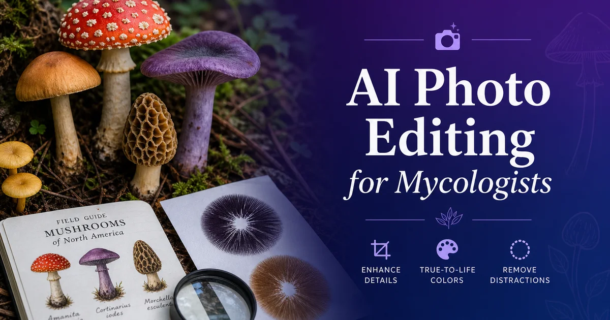
La mycologie — l'étude scientifique des champignons — dépend des archives visuelles plus lourdement que presque toute autre discipline biologique, car les caractères morphologiques qui distinguent une espèce fongique d'une autre sont souvent des textures de surface subtiles et des nuances de couleur précises. Des détails structurels tridimensionnels difficiles à décrire avec des mots mais immédiatement apparents sur une photographie bien réalisée. Un mycologue qui examine un champignon sur le terrain évalue la forme et la texture du chapeau, l'espacement et le type d'insertion des lames, le caractère de la surface du pied, les restes de voile, la couleur de la sporée, la couleur de la chair et toute réaction au meurtrissement, l'odeur, l'habitat et le substrat. Et la photographie qu'il prend doit capter suffisamment de ces caractères pour permettre une révision de l'identification plus tard, à un bureau ou par des collègues qui ne peuvent pas examiner le spécimen physique. Pour les mycologues experts, les contributeurs de science citoyenne, les auteurs de guides de terrain et la communauté grandissante des passionnés amateurs, la photographie est le pont entre l'observation sur le terrain et une identification fiable.
Les défis techniques de la photographie de champignons sont redoutables et propres au sujet. Les champignons poussent dans les environnements les plus difficiles à photographier que l'on puisse imaginer. Ombre profonde de la forêt, troncs en décomposition sous une lumière tachetée, litière de feuilles aux textures visuelles complexes, et sols où les spécimens bruns se fondent dans des arrière-plans bruns. Les spécimens eux-mêmes posent des défis optiques qui mettent à l'épreuve les systèmes d'appareils photo : les lames sont des structures parallèles délicates qui exigent un angle d'éclairage précis pour être résolues, les surfaces des chapeaux vont de revêtements visqueux brillants comme un miroir à des textures sèches et mates qui absorbent la lumière différemment, les couleurs couvrent tout le spectre visible et incluent souvent des nuances subtiles que les appareils peinent à reproduire fidèlement. La forme tridimensionnelle d'un champignon change le besoin d'éclairage d'un angle à l'autre. La plupart des mycologues de terrain travaillent avec l'appareil de leur téléphone sous la lumière disponible, produisant des images qui captent l'allure générale d'un spécimen mais perdent le détail fin nécessaire à une identification confiante au niveau de l'espèce.
Les outils d'édition photo par IA répondent aux défis spécifiques d'archives visuelles que les mycologues rencontrent à chaque étape de leur flux de travail. La suppression de l'arrière-plan isole les spécimens du sol forestier visuellement chaotique, permettant un affichage net dans les guides de terrain, les bases de données d'identification et les publications de recherche. L'amélioration par IA récupère le détail des lames, les textures de surface et les nuances de couleur que les limites des appareils et les conditions d'éclairage de terrain dégradent. La suppression d'objets nettoie les débris, les dégâts d'insectes et les artefacts de manipulation qui sont inévitables dans la photographie de spécimens de terrain. Ce guide couvre l'ensemble du flux de travail de photographie et d'édition mycologique, des techniques de collecte sur le terrain aux applications spécialisées comme les archives de sporées et la préparation d'images de microscopie, en mettant l'accent sur les caractères diagnostiques qui rendent la photographie de champignons scientifiquement précieuse plutôt que simplement décorative.
- Background Eraser isole les spécimens fongiques d'environnements de sol forestier complexes, permettant des planches taxonomiques nettes, des illustrations de guides de terrain et des images prêtes pour les bases de données avec des arrière-plans standardisés.
- AI Enhance récupère les détails morphologiques diagnostiques — espacement des lames, texture de surface du chapeau, restes de voile et caractère du pied — que les appareils de téléphone perdent dans l'éclairage difficile de l'ombre forestière.
- La préservation de la fidélité des couleurs durant l'amélioration est cruciale en mycologie, où de subtiles différences de teinte entre le chapeau, les lames et la chair peuvent distinguer une espèce comestible d'un sosie toxique dangereux.
- Magic Eraser supprime les complications de la photographie de terrain, y compris les débris de terre, les dégâts d'insectes, les traces de limaces, les fragments de feuilles et les artefacts de manipulation qui masquent les caractères diagnostiques du spécimen.
- L'exportation par lots produit des images au format spécifique pour les publications taxonomiques, les bases de données d'herbiers, les plateformes de science citoyenne et l'impression de guides de terrain. Des présentations éducatives à partir d'un seul fichier maître édité.
Technique de photographie de terrain : capturer les caractéristiques diagnostiques pour une identification fiable
Le fondement d'une photographie mycologique utile est une approche systématique pour capter chaque caractère diagnostique qu'offre un spécimen. Le caractère qui scelle une identification n'est souvent pas celui auquel on s'attend en appuyant sur le déclencheur. Les mycologues experts et les scientifiques citoyens expérimentés développent un protocole de prise de vue standardisé pour chaque récolte : un cliché de l'habitat montrant le spécimen dans son contexte de croissance et son type de substrat, une vue du dessus du chapeau révélant la texture de surface et le motif de couleur, une vue de dessous montrant le type d'hyménium (lames, pores, aiguillons ou surface lisse) et son insertion sur le pied, un cliché de profil complet montrant les proportions générales et tout reste de voile. Une coupe transversale révélant la couleur de la chair, son épaisseur et toute réaction de meurtrissure ou de changement de couleur survenant au contact de l'air. Ce protocole multi-angle garantit que les caractères essentiels à l'identification sont documentés même lorsque le photographe ne sait pas encore quels caractères se révéleront diagnostiques pour l'espèce particulière.
La gestion de l'éclairage sur le terrain est le facteur technique le plus déterminant en photographie de champignons. La différence entre des spécimens utiles et inutilisables se résume souvent à l'angle de la lumière. Le principe crucial est que les structures tridimensionnelles comme les lames, les pores et les textures de surface deviennent visibles grâce à l'ombre. Et l'ombre exige une lumière directionnelle inclinée par rapport à la surface du sujet. Une lumière directe venant du dessus, qu'elle provienne du soleil de midi ou du flash de l'appareil, éclaire toutes les surfaces de manière égale et élimine les ombres qui révèlent texture et structure. La technique de terrain la plus efficace consiste à se positionner de sorte que la lumière disponible frappe le spécimen de côté ou légèrement par l'arrière, créant des ombres qui révèlent les arêtes des lames, les bosses de surface, les textures fibrilleuses. La forme volumétrique du chapeau et du pied. Un petit carton réflecteur — même une page blanche de carnet — renvoyé vers le côté à l'ombre empêche le contraste de devenir trop extrême pour le capteur de l'appareil.
La photographie de sporée est une tâche d'archivage spécialisée que de nombreux mycologues trouvent étonnamment difficile à bien exécuter. Une sporée se réalise en posant un chapeau de champignon lames vers le bas sur du papier (moitié blanc, moitié sombre) pendant plusieurs heures, ce qui permet d'évaluer la couleur du dépôt de spores. Un caractère d'identification crucial pour de nombreux genres. La sporée obtenue est un dépôt délicat de spores microscopiques pouvant aller du blanc au crème, rose, brun, orange rouille, brun pourpré, jusqu'au noir de jais. Photographier les sporées exige un éclairage parfaitement uniforme sans reflets spéculaires sur la surface du papier, une gestion soigneuse des couleurs pour capter la véritable teinte de la sporée. Une résolution suffisante pour montrer le motif de la sporée correspondant à la disposition des lames. Les appareils de téléphone ont tendance à surexposer les sporées blanches en blanc pur et à comprimer les sporées sombres en un noir sans détail, perdant l'information de couleur subtile dont les mycologues ont besoin.
- Un protocole de prise de vue multi-angle standardisé — habitat, dessus du chapeau, dessous des lames, profil. Coupe transversale — garantit que tous les caractères diagnostiques sont documentés, quel que soit celui qui se révèle critique pour l'identification.
- Un éclairage latéral directionnel est essentiel pour révéler les structures tridimensionnelles des lames, les textures de surface et les restes de voile qui disparaissent sous un éclairage plat venu du dessus ou sous le flash de l'appareil.
- Un carton réflecteur blanc renvoyé vers le côté à l'ombre d'un spécimen éclairé en lumière directionnelle empêche le contraste excessif qui sature les capteurs des appareils de téléphone dans les conditions d'ombre forestière.
- La photographie de sporée exige un éclairage uniforme sans ombres, une reproduction fidèle des couleurs. Une résolution suffisante pour capter les subtiles différences de teinte entre le blanc, le crème, le rose et d'autres dépôts de couleur claire.
Suppression d'arrière-plan et isolation de spécimens pour la documentation taxonomique
La complexité visuelle des habitats fongiques naturels est le principal obstacle à des archives de spécimens nettes. Background Eraser s'y attaque directement en isolant le spécimen de son environnement. Un champignon poussant dans la litière de feuilles d'un sol forestier existe dans un champ visuel de brun sur brun sur brun. Feuilles mortes, terre, brindilles, autres matières organiques en décomposition — ce qui rend difficile pour le spectateur de distinguer les bords du spécimen du substrat, même lorsqu'il sait ce qu'il regarde. Le problème s'intensifie avec les espèces dont la coloration brune ou fauve correspond étroitement à leur substrat de croissance, les petits spécimens poussant en touffes où les carpophores individuels se chevauchent. Les spécimens poussant sous des troncs ou des branches, où le substrat de croissance lui-même devient un élément visuel déroutant. La suppression de l'arrière-plan sépare proprement le spécimen de tout ce bruit environnemental.
Le choix de l'arrière-plan de remplacement dépend de l'objectif d'archivage et doit suivre les conventions établies dans l'édition mycologique. Les planches taxonomiques et les descriptions formelles d'espèces ont par le passé utilisé des arrière-plans blancs ou gris très clair qui maximisent la fidélité des couleurs et permettent de comparer les caractères diagnostiques d'une planche à l'autre sans variation d'arrière-plan. Les illustrations de guides de terrain utilisent souvent un arrière-plan naturel simplifié. Une suggestion de texture forestière sans la complexité visuelle particulière du site de récolte réel — ce qui fournit un contexte sans distraction. Les bases de données et applications d'identification numériques bénéficient d'arrière-plans transparents qui permettent de composer les spécimens avec des étiquettes diagnostiques, des annotations de mesure et des images de comparaison. Pour chaque objectif, le remplacement d'arrière-plan par IA produit des résultats cohérents et standardisés qui exigeraient un studio portable pour être obtenus sur le terrain.
Conserver le mycélium basal, le point d'attache au substrat. Tout rhizomorphe ou cordon mycélien relié à la base du spécimen est important lorsqu'on isole des spécimens de champignons, car ces caractères portent une information diagnostique. Certaines espèces sont identifiées en partie par la façon dont la base du pied se rattache au sol. Qu'elle se termine par un bulbe, un sac de volve, un prolongement radicant ou un mycélium étalé. Background Eraser doit être appliqué de manière sélective ici, retirant le substrat environnant tout en conservant la terre ou le bois encore attaché à la base du pied qui révèle le mode de croissance. Cette approche sélective produit des images nettes et bien ciblées tout en conservant l'information morphologique à la jonction critique entre le carpophore fongique et le substrat de croissance.
- Les arrière-plans de sol forestier créent une complexité visuelle de brun sur brun qui rend les bords du spécimen difficiles à distinguer, surtout pour les espèces fauves ou brunes poussant sur des substrats assortis.
- Les planches taxonomiques utilisent des arrière-plans blancs pour la fidélité des couleurs, les guides de terrain utilisent des environnements naturels simplifiés pour le contexte, et les clés numériques utilisent des arrière-plans transparents pour la composition d'annotations.
- Le remplacement d'arrière-plan par IA produit des résultats standardisés sur des spécimens photographiés dans des lieux et conditions différents, égalant la cohérence d'un studio portable.
- La suppression sélective de l'arrière-plan préserve le mycélium basal, les restes de volve et les caractères d'attache au substrat à la base du pied qui portent une information d'identification diagnostique.
Améliorer les caractéristiques diagnostiques : détail des lamelles, texture de surface et fidélité des couleurs
La structure des lames est l'un des caractères diagnostiques les plus importants pour l'identification des champignons. C'est aussi l'un des plus difficiles à bien photographier dans les conditions de terrain. L'hyménium — la surface productrice de spores — se présente sous forme de plaques parallèles serrées (lames), de petits pores ronds, d'aiguillons ou d'épines pendants, ou de surfaces lisses à ridées selon l'espèce. Chaque type exige des stratégies d'amélioration différentes. Pour les champignons à lames, AI Enhance peut accentuer les arêtes individuelles des lames pour montrer le motif d'espacement (serrées ou espacées), le type d'insertion à la jonction du pied (libres, adnées, échancrées, décurrentes), le caractère de l'arête des lames (lisse, dentelée ou frangée). Les dégradés de couleur de l'arête des lames vers la chair du chapeau. L'amélioration doit accroître la séparation visible entre les plaques individuelles des lames sans créer d'artefacts d'arête artificiels qui pourraient être pris pour des caractères naturels. Une distinction clé entre l'amélioration à des fins artistiques et l'amélioration à des fins d'archivage scientifique.
La texture de surface du chapeau a un poids taxonomique majeur et varie énormément selon les genres fongiques. Les chapeaux visqueux ou glutineux ont un aspect humide et brillant même dans des conditions sèches, les chapeaux fibrilleux montrent des fibres radiales plaquées contre la surface, les chapeaux écailleux ont des motifs d'écailles saillantes distincts, les chapeaux lisses sont sans relief. Certaines espèces montrent des motifs distinctifs de craquelures ou d'aréolation en mûrissant. Les appareils de téléphone sous l'éclairage de terrain échouent souvent à résoudre ces caractères de surface, comprimant le brillant visqueux en un reflet uniforme et rendant la texture fibrilleuse comme lisse. L'amélioration par IA récupère ces détails de surface en augmentant le micro-contraste dans la plage de fréquences spécifique qui correspond aux caractères de texture de surface. L'amélioration doit rendre les textures visibles plus apparentes sans inventer de textures qui ne sont pas présentes. Là encore, la norme d'archivage scientifique consiste à révéler plutôt qu'à créer l'information visuelle.
La fidélité des couleurs en photographie mycologique n'est pas simplement esthétique — c'est une question de sécurité. De nombreux couples d'espèces comestible-toxique se distinguent principalement par des différences de couleur qui peuvent être subtiles en conditions de terrain : le chapeau jaune-orangé d'une girolle comestible face à l'orange-rouge légèrement plus prononcé d'un clitocybe lumineux toxique, les lames blanches d'une Amanita mortelle face aux lames d'abord roses puis brunes d'un Agaricus comestible, les tons olive du chapeau d'une amanite phalloïde face au blanc verdâtre d'un volvaire comestible. L'amélioration doit améliorer la visibilité et le détail des couleurs naturelles sans décaler les teintes d'une manière qui pourrait affecter l'identification. Cela signifie renforcer le contraste de luminance et la saturation dans des limites naturelles tout en évitant les algorithmes agressifs d'exaltation des couleurs qui font ressortir les paysages mais pourraient faire passer un champignon dangereux pour un champignon sûr.
- L'amélioration des lames accentue les arêtes individuelles des plaques pour révéler le motif d'espacement, le type d'insertion sur le pied, le caractère de l'arête. Les dégradés de couleur sans créer d'artefacts artificiels qui pourraient induire l'identification en erreur.
- L'amélioration de la texture de surface du chapeau récupère le brillant visqueux, les motifs de fibres fibrilleuses, les structures d'écailles et l'aréolation craquelée que les appareils de téléphone comprimaient en surfaces uniformes dans l'éclairage de terrain.
- La fidélité des couleurs est un besoin critique pour la sécurité. L'amélioration doit révéler les couleurs naturelles sans décalages de teinte qui pourraient masquer les subtiles différences distinguant les espèces comestibles de leurs sosies toxiques.
- L'amélioration pour la documentation scientifique suit une norme stricte consistant à révéler l'information visuelle existante plutôt qu'à créer ou exagérer des caractères qui ne sont pas présents sur le spécimen réel.
Applications spécialisées : sporées, microscopie et plateformes de science citoyenne
L'amélioration de sporée est une application spécialisée où les outils d'IA apportent une valeur majeure aux mycologues aux prises avec les limites techniques de la photographie de dépôts de spores délicats. La couleur subtile d'une sporée. Qui peut faire la différence entre des genres dans de nombreuses clés d'identification — est souvent perdue lorsque les appareils de téléphone calculent l'exposition automatique sur l'arrière-plan papier et réduisent la sporée à un blanc cramé ou à un sombre comprimé. L'amélioration par IA récupère la véritable couleur du dépôt de spores en normalisant l'exposition sur l'ensemble de l'image et en faisant ressortir la teinte subtile qui distingue le blanc du crème, le crème du jaune pâle, le jaune pâle du chamois. Tout le spectre à travers le rose, le saumon, la rouille, le brun, le brun pourpré et le noir. Pour les sporées mi-blanc mi-sombre réalisées sur un papier divisé, l'amélioration peut équilibrer l'exposition sur les deux moitiés afin que le même dépôt de spores soit visible sur les deux arrière-plans. C'est l'affichage standard pour les archives de sporées mycologiques.
L'édition d'images de microscopie étend la valeur des outils d'IA à la phase de laboratoire du travail mycologique. De nombreux caractères d'identification cruciaux — forme et ornementation des spores, morphologie des cystides, boucles d'anastomose sur les hyphes. Réactions de coloration chimique — ne sont visibles qu'au microscope, et les images capturées par des caméras d'oculaire de microscope ou par photographie au téléphone à travers l'oculaire souffrent souvent d'un éclairage inégal, d'aberration chromatique, d'une profondeur de champ limitée et de contamination de l'arrière-plan par des bulles et des débris du milieu de montage. Magic Eraser supprime les artefacts de montage, les débris d'arrière-plan et les éléments flous qui détournent l'attention des structures cibles. AI Enhance accentue l'ornementation des spores, le détail des parois et le contraste de coloration qui révèlent les caractères taxonomiques. La suppression de l'arrière-plan isole des spores individuelles ou des structures hyphales pour la mesure et la comparaison avec des images de référence.
Les contributions de science citoyenne à des plateformes comme iNaturalist, Mushroom Observer. Les bases de données des sociétés mycologiques régionales représentent le plus grand volume de photographie fongique au monde, et la précision d'identification de ces efforts communautaires dépend directement de la qualité des images. Les photographies de terrain éditées par IA avec des arrière-plans nets, des caractères diagnostiques améliorés. Des couleurs fidèles reçoivent des identifications plus rapides et plus assurées de la part des membres experts de la communauté qui examinent les soumissions. L'approche d'archivage multi-angle standardisée décrite dans ce guide, combinée à l'édition par IA pour nettoyer les compromis inévitables de la photographie de terrain, produit des soumissions de science citoyenne qui apportent une véritable valeur scientifique à la surveillance de la biodiversité, à la cartographie de l'aire de répartition des espèces et au suivi phénologique. Pour les mycologues amateurs qui peuvent photographier des centaines de spécimens lors d'une saison productive, les flux de travail de traitement par lots rendent pratique le maintien de la qualité d'archivage sur l'ensemble de la collection.
- L'amélioration de sporée récupère la teinte diagnostique — du blanc au crème, rose, rouille, brun pourpré, jusqu'au noir — que l'exposition automatique et les limites du capteur comprimaient en extrêmes non informatifs.
- L'édition d'images de microscopie supprime les artefacts de montage, corrige l'éclairage inégal et accentue l'ornementation des spores et le détail des hyphes pour l'évaluation des caractères taxonomiques.
- Les soumissions aux plateformes de science citoyenne avec des caractères diagnostiques améliorés par IA reçoivent des identifications expertes plus rapides et plus assurées, augmentant la valeur scientifique des données communautaires sur la biodiversité.
- Les flux de travail de traitement par lots permettent aux mycologues amateurs de maintenir une qualité d'archivage cohérente sur des centaines de spécimens collectés au cours de saisons de terrain productives.
How to do it
- 1
Photograph fungi specimens with lighting that reveals diagnostic morphological features
Set up specimen photography with diffused natural or artificial light that reveals the key identification features mycologists depend on. Cap surface texture, gill spacing and attachment type, stipe surface character, and color variations across the fruiting body. Side lighting at a low angle is key for capturing the three-dimensional form of gills, pores, teeth. Other hymenium structures that are critical for genus-level identification. Photograph each specimen from multiple standardized angles: cap surface from above, gill or pore surface from below with the cap tilted, a full profile showing cap-to-stipe proportions. A cross-section cut revealing internal flesh color and any color-change reactions upon exposure to air.
- 2
Remove distracting field backgrounds for clean specimen documentation
Use Background Eraser to isolate fungal specimens from the visually complex forest floor, rotting wood, leaf litter, and soil settings where they grow. Field photography is key for capturing fresh specimens in their natural substrate context, but the surrounding debris, competing vegetation. Variable lighting create backgrounds that obscure diagnostic features in identification guides and research publications. Replace with neutral backgrounds appropriate to the documentation purpose. White or light gray for taxonomic plates, natural but simplified woodland settings for field guides and educational materials, or transparent backgrounds for digital identification keys where specimens need to be composited with diagnostic annotations.
- 3
Enhance surface textures, gill detail, and color accuracy with AI Enhance
Apply AI Enhance to recover the fine morphological details that phone cameras and even consumer DSLRs struggle to resolve in fungal specimens. Cap surface textures — viscid, dry, scaly, fibrillose, or smooth — are primary identification features that photograph poorly in uncontrolled field lighting. Gill spacing, whether gills are free, adnate, adnexed, or decurrent. The presence of partial or universal veil remnants on cap and stipe are all details that benefit from boost. Color accuracy is mainly important in mycology where subtle hue differences distinguish edible species from toxic lookalikes. Enhancement should improve clarity without shifting the specimen's true colors.
- 4
Remove debris, damage, and competing organisms with Magic Eraser
Use Magic Eraser to clean up the natural-world complications that affect field mycology photography. Soil particles clinging to the stipe base, insect damage to cap surfaces, slug trails across gills, leaf fragments stuck to viscid caps, competing moss and lichen growth on shared substrate, and the inevitable finger smudges from handling specimens during collection. These elements are part of the field experience but they obscure the diagnostic features that make a photograph useful for spotting reference, educational publication, or research records. Remove them selectively while keeping the natural character of the specimen itself.
- 5
Export specimen images for field guides, research databases, and citizen science platforms
Save edited specimen photographs at full resolution with embedded color profile for archival and research use, then create versions optimized for each distribution channel. Taxonomic publications and herbarium databases need high-resolution images with standardized backgrounds and scale references. Field guide publishers require consistent formatting across all species plates with controlled color reproduction. Citizen science platforms like iNaturalist and Mushroom Observer benefit from clean, well-lit specimen photos that improve community identification accuracy. Educational displays and social media outreach need web-optimized versions that load quickly while keeping the morphological detail that makes mycology photography scientifically useful.
Sources
- Photographing Fungi: A Best Practice Guide for Citizen Science — iNaturalist Community Guidelines
- Mycological Specimen Photography Standards for Herbarium Collections — MycoBank — International Mycological Association
- Digital Imaging for Fungal Taxonomy and Identification — National Center for Biotechnology Information