
Edição de fotos com IA para nematologistas — Magic Eraser
Como nematologistas usam a edição de fotos com IA para limpeza de imagens de microscopia, aprimoramento de características diagnósticas e pranchas prontas para publicação. Aumente a nitidez dos detalhes do estilete, remova artefatos de lâmina e padronize micrografias.

Product Marketing
Revisado por Magic Eraser Editorial ·
Índice
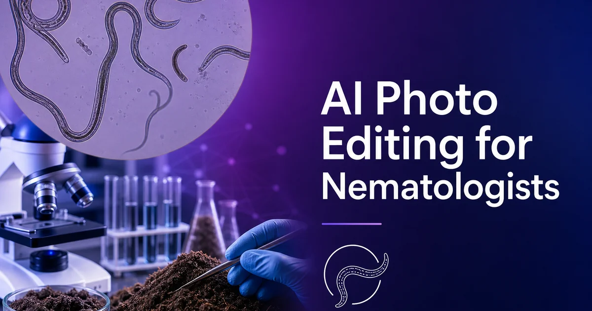
A nematologia — o estudo dos vermes cilíndricos (filo Nematoda) — é uma das disciplinas biológicas mais dependentes de imagem, porque os organismos que estuda são diagnosticados quase inteiramente por meio de caracteres morfológicos microscópicos. Com um milhão ou mais de espécies de nematoides estimadas na Terra e apenas cerca de 30.000 descritas atualmente, a capacidade de produzir com eficiência micrografias claras e detalhadas é fundamental para o trabalho taxonômico e diagnóstico que constitui o núcleo do campo. Cada descrição de espécie exige pranchas que mostrem as estruturas diagnósticas, cada relatório de diagnóstico agrícola exige registros fotográficos da espécie praga identificada. Cada levantamento ecológico precisa de registros de imagem da composição da comunidade de nematoides.
Os desafios fotográficos em nematologia têm raízes nos próprios organismos. Os nematoides são pequenos — a maioria das espécies relevantes para a agricultura e a medicina mede entre 0,5 e 5 milímetros de comprimento — e seus corpos são em grande parte transparentes, exigindo técnicas ópticas especializadas como o contraste de interferência diferencial (DIC) ou a microscopia de contraste de fase para visualizar as estruturas internas. Os caracteres diagnósticos que distinguem as espécies muitas vezes são medidos em micrômetros: o comprimento e a forma do estilete (uma estrutura de alimentação nos nematoides fitoparasitas), a disposição das glândulas faríngeas, a forma das espículas nos machos. A posição e a morfologia da vulva nas fêmeas. Captar esses caracteres com nitidez suficiente para uma identificação confiável é tecnicamente exigente.
As ferramentas de edição de fotos com IA enfrentam os desafios específicos de pós-processamento da microscopia de nematoides. A limpeza de fundo remove os detritos, as bolhas de ar e os organismos coocorrentes que contaminam as preparações em lâmina. O realce de detalhes recupera os finos caracteres estruturais. Os padrões de anelação cuticular, as formas dos botões do estilete, a morfologia das válvulas faríngeas — que são diagnosticamente cruciais, mas frequentemente indistintos nas micrografias padrão. A normalização de imagens uniformiza as micrografias obtidas em diferentes sistemas de microscópio e sessões de imageamento para um trabalho comparativo coerente. Para os nematologistas que processam centenas de micrografias por estudo taxonômico, um pós-processamento eficiente assistido por IA acelera diretamente o ritmo de descrição de espécies e de elaboração de relatórios diagnósticos.
- A remoção de fundo elimina os detritos da lâmina, as bolhas de ar e os artefatos do meio de montagem sem alterar o contorno do corpo do nematoide nem as estruturas internas.
- O AI Enhance afina as estruturas diagnosticamente cruciais — morfologia do estilete, disposição faríngea, forma vulvar e forma das espículas — medidas em micrômetros.
- O Magic Eraser remove os organismos coocorrentes e a contaminação particulada das preparações em lâmina, preservando a integridade do espécime alvo.
- A normalização de imagens uniformiza as micrografias entre os sistemas DIC, de contraste de fase e de campo claro para uma comparação lado a lado sem viés.
- As pranchas prontas para publicação a 300 DPI com barras de escala e rotulagem coerentes atendem aos requisitos das revistas para descrições taxonômicas.
Limpeza de imagens de microscopia para preparações de lâminas de nematoides
As preparações de nematoides em lâmina são ambientes intrinsecamente desordenados. O processo padrão envolve matar o nematoide com calor brando, fixá-lo em solução de formol ou TAF, clarificá-lo para aumentar a transparência (muitas vezes com glicerina ou lactofenol). Montá-lo em uma lâmina de vidro sob uma lamínula. Em cada etapa desse processo, artefatos podem ser introduzidos. Bolhas de ar aprisionadas durante a montagem, produtos químicos de fixação precipitados, detritos do solo ou do tecido vegetal de onde o nematoide foi extraído, bactérias e esporos fúngicos que colonizam o meio de montagem, e arranhões ou impressões digitais nas superfícies da lâmina e da lamínula.
Esses artefatos são muito mais do que problemas estéticos. As bolhas de ar e as partículas de detritos podem ocultar justamente as estruturas que os nematologistas precisam examinar para a identificação. Uma bolha posicionada sobre a região faríngea esconde a disposição das glândulas faríngeas que distingue os gêneros. Uma partícula de detrito sobre a extremidade da cauda oculta a forma que separa as espécies. No fluxo de trabalho microscópico convencional, o nematologista simplesmente refoca através ou ao redor desses obstáculos. Na fotomicrografia, o artefato fica registrado permanentemente na imagem e deve ser tratado no pós-processamento.
As ferramentas de remoção baseadas em IA lidam com esses artefatos com a precisão que a microscopia de nematoides exige. O Magic Eraser pode remover uma bolha de ar sobre a região faríngea, preenchendo a área com o fundo óptico e as estruturas do nematoide visíveis na área circundante. Pode eliminar partículas de detritos do meio de montagem sem alterar o contorno do corpo do nematoide nem o padrão de anelação cuticular do espécime subjacente. O requisito chave é que a remoção não deve inventar nem alterar qualquer estrutura morfológica. A área preenchida deve ser uma extensão fiel das estruturas reais visíveis ao lado do artefato, não uma alucinação da IA sobre o que poderia estar por baixo.
- A preparação da lâmina introduz bolhas de ar, precipitados de fixação, detritos de solo, contaminação microbiana e danos à superfície do vidro.
- Os artefatos podem ocultar estruturas diagnosticamente cruciais — glândulas faríngeas, extremidades da cauda, estruturas reprodutivas — nas micrografias capturadas.
- A remoção por IA preenche as áreas de artefatos com um fundo e uma estrutura do espécime opticamente coerentes a partir do contexto circundante.
- A integridade morfológica é a restrição crítica — a remoção não deve inventar, alterar nem reposicionar qualquer estrutura de nematoide visível na imagem.
Aprimorando características diagnósticas para identificação de espécies
A identificação de espécies de nematoides depende de um conjunto específico de caracteres morfológicos e morfométricos que devem ser claramente visíveis e mensuráveis nas micrografias. Para os nematoides fitoparasitas, o estilete — uma estrutura de alimentação oca e protrátil na cabeça — costuma ser o primeiro caráter diagnóstico examinado. O comprimento do estilete, a largura da haste, a forma e o tamanho dos botões basais (redondos, voltados para a frente, inclinados para trás). As proporções gerais distinguem gêneros e espécies dentro dos principais grupos fitoparasitas. Essas diferenças podem ser tão pequenas quanto dois a três micrômetros no comprimento total do estilete entre espécies estreitamente aparentadas, exigindo a máxima nitidez de imagem para medi-las de forma confiável.
O AI Enhance aplicado às micrografias de nematoides aumenta o contraste local dentro da parede corporal transparente e das estruturas internas sem amplificar o ruído de fundo nem os artefatos de microscopia. A região faríngea — contendo o metacorpo, o istmo, as glândulas faríngeas. E seus núcleos associados — é uma estrutura tridimensional complexa que deve ser imageada através da profundidade do corpo do nematoide. Um realce que aumenta o contraste entre essas estruturas internas sobrepostas as torna distinguíveis uma a uma em uma micrografia bidimensional. É crucial para determinar a disposição e as posições relativas das glândulas faríngeas que caracterizam os diferentes grupos de nematoides.
As estruturas reprodutivas fornecem outro conjunto-chave de caracteres. Nas fêmeas, a posição vulvar (expressa como porcentagem do comprimento corporal total a partir da extremidade anterior), a forma e a estrutura da vagina. A disposição dos ovários (pareados ou únicos, distendidos ou refletidos) são caracteres diagnósticos padrão. Nos machos, a forma, o comprimento e a curvatura das espículas, junto com a morfologia do gubernáculo e quaisquer asas caudais, são usados para distinguir as espécies. O AI Enhance, que afina as bordas dessas estruturas de tecido mole e aumenta seu contraste em relação ao fluido da cavidade corporal circundante, as torna mensuráveis e descritíveis em micrografias onde, de outro modo, apareceriam apenas como vagas sombras internas.
- As dimensões do estilete distinguem gêneros e espécies — diferenças de dois a três micrômetros no comprimento total separam táxons estreitamente aparentados.
- A disposição e a posição das glândulas faríngeas exigem realce de contraste para resolver estruturas tridimensionais sobrepostas em micrografias 2D.
- Os caracteres reprodutivos femininos — posição vulvar, estrutura vaginal, disposição dos ovários — exigem uma definição nítida das bordas dos tecidos moles.
- A forma e a curvatura das espículas masculinas, e a morfologia do gubernáculo, são afinadas de vagas sombras internas a estruturas diagnósticas mensuráveis.
Padronizando micrografias entre plataformas de microscopia
Os nematologistas que trabalham em várias instituições, que colaboram em taxonomia revisional ou que comparam seus espécimes com descrições publicadas precisam lidar com a realidade de que as micrografias de diferentes sistemas de microscópio parecem diferentes mesmo quando mostram as mesmas estruturas. A microscopia DIC produz imagens com um característico efeito de relevo pseudotridimensional e uma dominante de cor (muitas vezes castanho-dourado ou cinza-azulado) que varia entre fabricantes de microscópios e configurações do prisma DIC. A microscopia de contraste de fase produz halos brilhantes ao redor das bordas do espécime e um brilho de fundo que depende do alinhamento do anel de fase e das configurações do condensador. A microscopia de campo claro padrão produz imagens planas e de baixo contraste de espécimes transparentes que dependem inteiramente da clarificação e da coloração do espécime para sua visibilidade.
As ferramentas de normalização com IA uniformizam essas diferenças ópticas para que as micrografias de diferentes sistemas possam ser comparadas sem viés. A normalização de cor remove as dominantes de cor específicas do fabricante dos sistemas DIC, produzindo imagens com fundos neutros onde a única cor provém do próprio espécime. A redução de halos nas imagens de contraste de fase remove os artefatos de borda brilhante que podem ocultar finos caracteres cuticulares e tornar imprecisas as medições do contorno corporal. A normalização do brilho de fundo garante que o contraste entre o espécime e o fundo seja coerente entre as imagens, evitando a ilusão visual em que um nematoide parece mais escuro ou mais grosso em uma imagem simplesmente porque o fundo é mais claro.
Para as publicações colaborativas de vários autores e os grupos de trabalho internacionais sobre taxonomia de nematoides, essa normalização é sobretudo valiosa. Uma revisão de um gênero de nematoides pode incluir micrografias contribuídas por pesquisadores de dez instituições diferentes, cada um usando equipamentos de microscópio distintos. Sem normalização, as pranchas parecem heterogêneas. Dominantes de cor diferentes, níveis de contraste diferentes, tons de fundo diferentes — desviando o leitor das comparações morfológicas que as figuras pretendem apoiar. As imagens normalizadas criam pranchas visualmente coerentes em que a atenção do leitor é dirigida às diferenças reais entre as espécies, em vez das diferenças entre os microscópios.
- Os sistemas DIC, de contraste de fase e de campo claro produzem micrografias visualmente distintas das mesmas estruturas de nematoides devido aos seus princípios ópticos.
- A normalização de cor remove as dominantes de cor DIC específicas do fabricante, produzindo fundos neutros onde resta apenas a cor do espécime.
- A redução de halos do contraste de fase remove os artefatos de borda brilhante que ocultam caracteres cuticulares e distorcem as medições do contorno corporal.
- As publicações colaborativas multi-institucionais exigem micrografias normalizadas para pranchas visualmente coerentes que apoiem a comparação morfológica.
Diagnósticos agrícolas e fluxos de trabalho de identificação rápida
A nematologia agrícola opera sob uma pressão de tempo que a taxonomia acadêmica não enfrenta. Quando um agricultor envia uma amostra de solo ou uma cultura mostra sintomas de dano por nematoides, o laboratório de diagnóstico deve identificar as espécies de nematoides presentes e fornecer recomendações de manejo dentro de um prazo que permita ao produtor agir — muitas vezes dias, não meses. Extração, montagem, microscopia e identificação rápidas são práticas padrão nos laboratórios de diagnóstico agrícola, e os registros fotográficos de cada identificação servem tanto como documento de controle de qualidade quanto como referência para identificações futuras.
A edição de fotos com IA acelera a etapa de documentação dos fluxos de trabalho diagnósticos. Um técnico pode fotografar os caracteres diagnósticos em cada ampliação, processar em lote as micrografias por meio da limpeza de fundo e do realce. Produzir um documento diagnóstico limpo em minutos, em vez das horas que o pós-processamento manual em um software de edição de imagens convencional exigiria. Essa aceleração é sobretudo valiosa durante os picos de submissão. A primavera e o outono nas regiões temperadas — quando os laboratórios de diagnóstico podem processar dezenas de amostras por dia e os registros fotográficos podem se tornar um gargalo se cada imagem exigir atenção individual.
Para os materiais de treinamento e referência diagnósticos, as micrografias realçadas por IA melhoram enormemente a qualidade dos guias de identificação e dos recursos de treinamento. Uma chave de diagnóstico ilustrada com micrografias claras e realçadas que mostram os caracteres distintivos cruciais de cada espécie é mais útil do que uma ilustrada com micrografias brutas onde os detalhes diagnósticos estão ocultos por artefatos e baixo contraste. Muitos laboratórios de diagnóstico mantêm bibliotecas internas de imagens de referência das espécies comumente encontradas em sua região. O AI Enhance aplicado retrospectivamente às imagens mais antigas dessas bibliotecas pode elevar toda a coleção a um padrão de qualidade coerente sem refotografar cada lâmina.
- Os diagnósticos agrícolas exigem identificação rápida e documentação fotográfica em dias para recomendações acionáveis ao produtor.
- O processamento em lote por IA das micrografias diagnósticas reduz o pós-processamento por imagem de minutos para segundos durante os picos de submissão.
- Os materiais de treinamento e referência beneficiam-se de micrografias realçadas que mostram claramente os caracteres distintivos de cada espécie diagnóstica.
- O realce retrospectivo por IA eleva as bibliotecas históricas de imagens de referência a uma qualidade coerente sem refotografar as lâminas arquivadas.
How to do it
- 1
Capture nematode specimens with standardized microscopy setups
Mount nematodes on glass slides using right fixation and clearing techniques for the structures you need to document. Use differential interference contrast (DIC) or phase contrast microscopy to visualize the transparent nematode body. Photograph at standardized magnifications — often 100x for whole-body habitus images and 400x to 1000x for diagnostic features like the head, stylet, pharynx, vulva, and tail region. Include a calibrated scale bar in every micrograph.
- 2
Remove background artifacts and mounting medium debris
Use Magic Eraser to remove air bubbles trapped in the mounting medium, debris particles from the fixation process, glass slide scratches, cover slip edges visible in the frame. Any co-occurring organisms that are not the target specimen. Nematode slide preparations frequently contain particulate matter that obscures fine morphological details. Remove these distractions without altering the nematode body outline, internal structures, or cuticular features.
- 3
Enhance diagnostic morphological features for identification
Use AI Enhance to sharpen the fine structures that drive nematode spotting. Stylet morphology and basal knob shape, pharyngeal gland arrangement, excretory pore position, vulval structure, spicule shape in males, and tail terminus form. Many of these features are measured in micrometers and require maximum image clarity to distinguish between closely related species. Boost should increase local contrast within the nematode body without introducing artifacts at structural boundaries.
- 4
Normalize microscopy images taken with different optical setups
Use AI color correction to standardize images captured across different microscope systems, illumination sources, and optical configurations. DIC images have a trait color cast that varies between microscope brands. Phase contrast images vary in background brightness and halo intensity. Normalize these differences so that specimens photographed on different systems can be compared side by side in publications, spotting guides. Diagnostic reports without color or contrast bias.
- 5
Prepare figure plates with multi-view diagnostic image sets
Arrange processed micrographs into standardized figure plates showing the complete diagnostic image set for species description. Whole-body habitus, head en face view, stylet detail, pharyngeal region, reproductive structures (vulva and ovary arrangement in females, spicule and gubernaculum in males), and tail region. Maintain consistent scale indicators and labeling conventions. Export at 300 DPI minimum for journal submission, with inset panels for magnified details of taxonomically critical structures.
Fontes
- Microscopy and Imaging Techniques for Nematode Identification — Nematology (Brill Academic Publishers)
- Society of Nematologists: Resources for Nematode Research — Society of Nematologists
- Interactive Diagnostic Key to Plant Parasitic and Soil Nematodes — University of Nebraska–Lincoln