
Edição de fotos com IA para liquenologistas: Documente líquens e biomonitoramento — Magic Eraser
Edição especializada de fotografia de líquens para liquenologistas e cientistas ambientais. Ferramentas de IA para registros de talo, detalhes de apotécios, digitalização de herbário e biomonitoramento da qualidade do ar.
SEO & Growth
Revisado por Magic Eraser Editorial ·
Índice
- Técnicas de fotografia de campo para documentação de líquens em substratos naturais
- Remoção de fundo e isolamento de espécimes de substratos naturais complexos
- Aprimorando características diagnósticas: apotécios, sorédios, isídios e testes químicos pontuais
- Fotografia de líquens para biomonitoramento, levantamentos ecológicos e conservação
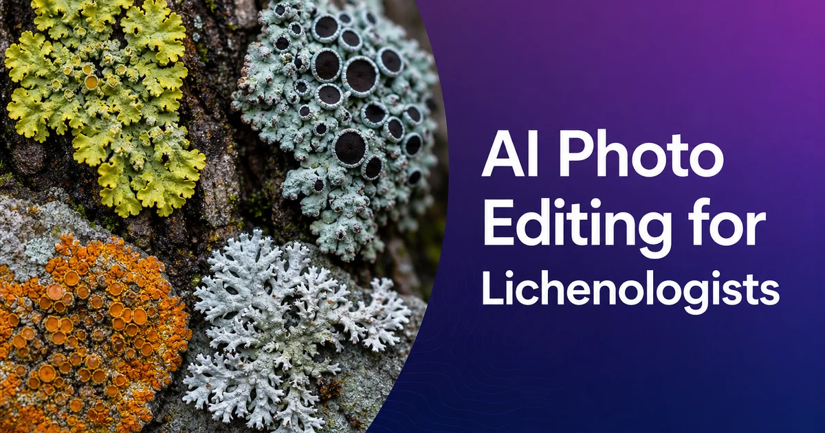
A liquenologia — o estudo dos líquenes, esses notáveis organismos simbióticos compostos por fungos e parceiros fotossintéticos — apoia-se em seu cerne nos registros visuais para a identificação de espécies e o monitoramento ecológico. E nos registros de herbário que ancoram a pesquisa taxonômica. Os líquenes são identificados por uma combinação de caracteres morfológicos e testes químicos pontuais. E, cada vez mais, pela análise do DNA, mas o primeiro nível de identificação e o mais acessível é o visual: a forma de crescimento do talo, a cor e a textura da superfície superior, a estrutura dos órgãos reprodutores e o porte geral do organismo sobre seu substrato. Esses caracteres vão de traços visíveis a olho nu. Seja o líquen crostoso, folioso ou fruticoso, e sua cor de conjunto — até detalhes microscópicos da estrutura dos esporos e da anatomia do córtex que exigem cortes e microscopia composta. A fotografia serve a toda a amplitude desse fluxo de trabalho de identificação, dos registros de campo ao detalhe macro até o arquivamento.
Os desafios fotográficos enfrentados pelos liquenólogos são singulares e exigentes. Os líquenes são pequenos: muitas espécies têm talos medidos em centímetros com traços diagnósticos medidos em frações de milímetro. Crescem sobre superfícies visualmente complexas que competem com o espécime pela atenção do observador: cascas rugosas com sua própria textura e variação de cor, paredões rochosos com padrões de grão mineral, muros antigos com juntas de argamassa e manchas de intemperismo. Superfícies de solo com musgo, serapilheira e outros organismos. Os próprios líquenes têm, com frequência, cores sutis. Tons cinza-esverdeados, oliva e marrons que se fundem com seus substratos em vez de contrastar com eles. E os traços diagnósticos mais importantes. A cor do disco apotecial, a textura soredial, a forma isidial e a estrutura da superfície do córtex — são precisamente os traços que as câmeras de telefone têm dificuldade de resolver, porque existem na escala em que o tamanho do pixel do sensor, a difração da lente e a precisão do autofoco se tornam todos fatores limitantes.
As ferramentas de edição de fotos com IA enfrentam esses desafios liquenológicos por meio de um fluxo de trabalho concebido para o registro de espécimes em todas as escalas. O Background Eraser isola os líquenes do ruído visual de seus substratos, criando imagens limpas que apresentam a forma do talo e a definição das margens com a nitidez dos espécimes de herbário prensados, mantendo ao mesmo tempo a estrutura tridimensional que prensar um líquen. Se é que se consegue sequer destacá-lo de seu substrato — com o tempo destrói. O AI Enhance recupera o detalhe diagnóstico em apotécios, soródios, isídios e na textura de superfície que as câmeras de telefone achatam em manchas sem traços. O Magic Eraser remove a contaminação ambiental. Películas de água, detritos caídos, organismos concorrentes — que compromete as fotografias de campo para fins de registro. Este guia cobre o fluxo de trabalho completo para liquenólogos, das técnicas de fotografia de campo à edição para fins de identificação e arquivamento, até a exportação para bancos de dados de herbário, relatórios de biomonitoramento e publicações.
- O Background Eraser isola os espécimes de líquenes de substratos visualmente complexos — casca, rocha, argamassa, solo — criando documentação de qualidade de herbário que preserva a morfologia tridimensional do talo.
- O AI Enhance recupera a cor do disco apotecial, a textura soredial e a forma isidial. O detalhe da superfície do córtex que as câmeras de telefone não conseguem resolver em organismos medidos em centímetros com traços em frações de milímetro.
- O Magic Eraser remove as películas de água que alteram a cor aparente do talo, os detritos caídos, os organismos concorrentes e as manchas indesejadas de testes químicos pontuais do substrato circundante.
- Uma edição coerente ao longo dos levantamentos de comunidades de líquenes garante uma qualidade de documentação padronizada para os relatórios de biomonitoramento da qualidade do ar e os programas de monitoramento ecológico de longo prazo.
- A exportação em lote cria imagens dimensionadas para bancos de dados de herbário, chaves de identificação interativas, registros de biomonitoramento e publicação em periódicos liquenológicos a partir de fotografias-mestre únicas já editadas.
Técnicas de fotografia de campo para documentação de líquens em substratos naturais
A fotografia de líquenes apresenta um conjunto único de desafios que decorrem dos hábitos de crescimento dos organismos, do tamanho reduzido e da semelhança visual com seus substratos. Ao contrário da maioria dos organismos que os fotógrafos podem isolar contra um fundo contrastante, os líquenes estão intimamente ligados às superfícies que colonizam. Os líquenes crostosos estão literalmente incrustados em seu substrato, com apenas o córtex superior visível; os foliosos aderem por rizinas que se agarram à casca ou às superfícies rochosas; e mesmo os fruticosos fixam-se em um único ponto a partir do qual o talo ramificado se estende. Isso significa que a fotografia deve incluir o substrato, a menos que o espécime tenha sido coletado e levado a um estúdio. O substrato introduz uma complexidade visual que compete com o espécime pela atenção. A casca das árvores tem cor, textura e padrão próprios que podem suplantar os tons sutis de um líquen crostoso cinza-esverdeado. As superfícies rochosas têm grão mineral e padrões de intemperismo que criam ruído visual ao redor e por baixo do espécime. Gerir essa competição do substrato é o desafio primordial da fotografia de líquenes de campo.
A iluminação para a fotografia de líquenes deve revelar a textura da superfície sem criar as sombras duras que ocultam o detalhe em organismos de relevo muito baixo. Os líquenes crostosos podem projetar-se menos de um milímetro acima de seu substrato, o que significa que qualquer sombra lançada pelo próprio líquen é muito curta e facilmente perdida no padrão de sombras do próprio substrato. Os líquenes foliosos têm uma estrutura mais tridimensional, mas seus traços diagnósticos. A textura do córtex superior, a cor da medula visível através das fendas do córtex e a estrutura dos sorais e isídios — revelam-se melhor sob uma luz suave e uniforme que evita os reflexos especulares e as sombras profundas criados pela luz solar direta. Um difusor portátil — mesmo uma folha de papel branco mantida entre o sol e o espécime — pode transformar a luz direta dura na iluminação suave que revela o detalhe da superfície do líquen sem o contraste que o esconde.
O registro da escala é fundamental na fotografia de líquenes porque os espécimes vão de minúsculas manchas crostosas de poucos milímetros a grandes talos foliosos que se estendem por trinta centímetros ou mais. O tamanho é, em si, um caráter diagnóstico para alguns grupos de espécies. Uma pequena régua ou cartão de escala colocado ao lado do espécime na fotografia fornece a referência de tamanho que permite a observadores posteriores julgar as dimensões absolutas de apotécios, sorais e outros traços diagnósticos. Ao fotografar líquenes para levantamentos de biomonitoramento, uma abordagem coerente do posicionamento da escala garante que as fotografias de espécimes de diferentes datas de levantamento e de diferentes pesquisadores sejam diretamente comparáveis. Isso é fundamental para detectar as mudanças na composição da comunidade de líquenes que indicam variações na qualidade do ar ao longo do tempo.
- Os líquenes crostosos incrustados no substrato e os foliosos aderidos por rizinas não podem ser separados para a fotografia. O substrato é sempre parte da imagem, competindo visualmente com os traços diagnósticos.
- A iluminação suave e difusa revela a textura da superfície em organismos com relevo submilimétrico, evitando as sombras duras e os reflexos especulares que ocultam o detalhe diagnóstico do córtex e dos soródios.
- Os indicadores de escala ao lado dos espécimes fornecem a referência de tamanho absoluto necessária para a medição apotecial e a comparação entre levantamentos nos programas de biomonitoramento de longo prazo.
- Uma abordagem fotográfica coerente ao longo das datas de levantamento e entre pesquisadores garante uma documentação diretamente comparável para detectar mudanças na qualidade do ar por meio das variações na composição da comunidade de líquenes.
Remoção de fundo e isolamento de espécimes de substratos naturais complexos
Isolar um líquen de seu substrato em uma fotografia é tecnicamente desafiador, porque o limite entre organismo e substrato costuma ser gradual em vez de nítido. Os líquenes crostosos, em particular, integram-se de modo tão completo com seu substrato que a margem do talo. O limite entre o líquen e o substrato nu ou os organismos adjacentes — pode ser definido apenas por uma sutil mudança de cor em vez de uma borda física. Os líquenes foliosos têm margens mais definidas, onde as pontas dos lobos se erguem ligeiramente do substrato, criando uma estreita linha de sombra que a detecção de bordas por IA pode seguir. Os líquenes fruticosos têm os limites mais nítidos, pois suas estruturas ramificadas se afastam do substrato. Têm também os contornos mais complexos, com ramificação fina que exige um traçado preciso das bordas para preservar o padrão de ramificação diagnóstico e a morfologia dos lobos.
O Background Eraser lida com esses graus variáveis de definição de limite combinando a detecção de diferença de cor com um reconhecimento de padrões aprendido que distingue o tecido do líquen dos substratos comuns. A IA foi treinada nas assinaturas visuais das combinações líquen-sobre-casca, líquen-sobre-rocha. Líquen-sobre-solo, o que lhe permite identificar as sutis transições de cor e textura que marcam as margens do talo, mesmo quando o contraste entre líquen e substrato é baixo. Para espécies crostosas em que o limite é genuinamente ambíguo, a detecção de bordas pode ser guiada ajustando-se a sensibilidade para favorecer a inclusão das áreas limítrofes (mantendo o talo completo ao custo de incluir algum substrato) ou a exclusão (produzindo um recorte mais limpo ao risco de aparar a margem do talo).
Substituir o fundo do substrato por tons neutros cria imagens de espécimes adequadas aos registros de bancos de dados de herbário e às ilustrações de chaves de identificação. À comparação lado a lado de espécimes de diferentes substratos e localidades. Essa padronização é especialmente valiosa em liquenologia porque a mesma espécie pode parecer marcadamente diferente em substratos distintos. Um líquen que cresce na casca clara de uma bétula aparece mais escuro do que a mesma espécie na casca escura de um carvalho, porque a cor percebida é influenciada pelo contraste simultâneo com o tom do substrato circundante. Isolar os espécimes sobre fundos neutros idênticos elimina essa distorção de cor dependente do substrato, permitindo comparar diretamente a cor real do talo entre espécimes coletados de árvores, rochas ou outras superfícies diferentes.
- As margens dos líquenes crostosos definidas apenas por sutis mudanças de cor exigem um reconhecimento de padrões por IA treinado em combinações líquen-substrato para identificar os limites onde as bordas físicas estão ausentes.
- As pontas dos lobos foliosos criam estreitas linhas de sombra que a detecção de bordas pode seguir, enquanto as estruturas ramificadas fruticosas precisam de um traçado preciso para preservar o padrão de ramificação diagnóstico e a morfologia.
- O ajuste de sensibilidade equilibra a inclusão completa do talo com uma definição de bordas limpa para espécimes cujo limite organismo-substrato é genuinamente ambíguo.
- A padronização com fundo neutro elimina o contraste simultâneo dependente do substrato que faz espécies idênticas parecerem de cores diferentes na casca clara de uma bétula em comparação com a casca escura de um carvalho.
Aprimorando características diagnósticas: apotécios, sorédios, isídios e testes químicos pontuais
Os apotécios — os corpos de frutificação em forma de taça ou de disco que produzem os esporos do líquen — estão entre os traços diagnósticos mais importantes para a identificação ao nível de espécie. Seu fino detalhe estrutural é precisamente o que o reforço por IA recupera melhor das fotografias de campo. A cor do disco apotecial (que varia do preto ao marrom, laranja, rosa. Vermelho até o bege pálido conforme a espécie), a estrutura da margem apotecial (se corresponde à cor do talo, indicando um tipo lecanorino, ou se é da mesma cor que o disco, indicando um tipo lecideíno) e a distribuição e densidade dos apotécios ao longo do talo fornecem todos caracteres de identificação que liquenólogos experientes leem num relance em boas fotografias, mas não conseguem extrair das imagens de campo subexpostas e de baixa resolução que as câmeras de telefone costumam produzir nos habitats sombreados onde cresce a maioria dos líquenes.
As estruturas reprodutivas vegetativas — soródios e isídios — exigem uma resolução efetiva ainda maior, porque costumam ser menores que os apotécios e seu valor diagnóstico reside na forma e na textura de superfície, mais do que apenas na cor e distribuição. Os soródios são grânulos pulverulentos de hifas fúngicas envolvendo células de algas, produzidos em estruturas chamadas sorais que podem ser puntiformes, lineares, labriformes ou difusos pela superfície do talo. Os isídios são excrescências digitiformes, cilíndricas ou coralóides da superfície do talo que se desprendem para a dispersão. A distinção entre soródios e isídios. E entre os diferentes tipos de cada um — é um caráter de identificação primário em grandes gêneros como Parmelia, Usnea e Cladonia. O AI Enhance aumenta o microcontraste que torna visível a diferença de textura entre a superfície granular dos soródios e a superfície corticada lisa dos isídios em fotografias tiradas no limite de resolução das lentes macro de telefone.
Os testes químicos pontuais são um traço distintivo da identificação de líquenes que cria necessidades fotográficas específicas. Os liquenólogos aplicam gotas de reagentes químicos. Hidróxido de potássio (K), hipoclorito de sódio (C) e parafenilenodiamina (P ou PD) — diretamente sobre a superfície do talo e observam as reações de cor resultantes, que indicam a presença de metabólitos secundários específicos do líquen, taxonomicamente diagnósticos. Documentar fotograficamente essas reações de cor exige captar a mudança de cor, muitas vezes transitória, na pequena área onde o reagente foi aplicado, mostrando ao mesmo tempo talo circundante suficiente para identificar o espécime. O AI Enhance pode aumentar a visibilidade de reações de cor sutis. O Magic Eraser pode limpar o reagente que se espalhou sobre o substrato circundante sem afetar a área de reação no próprio talo.
- A cor do disco apotecial, a estrutura da margem (lecanorino versus lecideíno). O padrão de distribuição fornecem caracteres de identificação primários recuperáveis pelo reforço por IA de imagens de campo subexpostas.
- A distinção entre soródios e isídios — textura de superfície granular versus corticada — é um caráter de identificação primário nos gêneros principais que exige reforço de microcontraste para ser resolvido a partir de imagens macro de telefone.
- A documentação dos testes químicos pontuais capta as reações de cor transitórias dos reagentes K, C e PD que indicam metabólitos secundários taxonomicamente diagnósticos na superfície do talo.
- O Magic Eraser limpa o reagente espalhado sobre o substrato circundante mantendo a reação de cor diagnóstica no talo, combinando os registros dos caracteres de identificação tanto morfológicos quanto químicos.
Fotografia de líquens para biomonitoramento, levantamentos ecológicos e conservação
Além dos registros taxonômicos, os liquenólogos usam cada vez mais a fotografia em programas de monitoramento ecológico nos quais os líquenes servem de bioindicadores da qualidade do ar e da saúde ambiental. Os líquenes são excepcionalmente sensíveis aos poluentes atmosféricos. Sobretudo ao dióxido de enxofre e aos compostos de nitrogênio — porque absorvem água e gases dissolvidos diretamente pela superfície, sem a cutícula protetora que protege as plantas vasculares. As mudanças na composição da comunidade de líquenes em árvores ou rochas de levantamento padronizadas refletem as mudanças na qualidade do ar ao longo do tempo, tornando o monitoramento de líquenes um complemento econômico à medição instrumental da qualidade do ar. Os registros fotográficos das parcelas de levantamento fornecem o registro visual permanente que permite reavaliar e verificar a composição da comunidade de líquenes anos após o levantamento original, sustentando os programas de monitoramento de longo prazo que a avaliação da qualidade do ar exige.
A fotografia de levantamento padronizada exige coerência entre múltiplos pesquisadores e datas de levantamento. E condições ambientais para produzir registros comparáveis que sustentem a detecção de tendências. As ferramentas de edição com IA tornam essa padronização prática ao normalizar as variáveis que diferem entre as sessões de campo. As condições de iluminação mudam com o tempo e a estação, o equipamento fotográfico varia entre pesquisadores, os níveis de umidade do substrato alteram a cor do líquen entre visitas, e a serapilheira acumulada e o trânsito de invertebrados nas parcelas de levantamento diferem de uma sessão para outra. A limpeza do fundo, a normalização da exposição por reforço e a remoção de elementos ambientais transitórios criam um conjunto de fotografias de levantamento em que as únicas diferenças refletem mudanças genuínas na comunidade de líquenes em vez de variação acidental nas condições de fotografia.
As aplicações de conservação da fotografia de líquenes incluem documentar espécies raras e ameaçadas para avaliações de estado de conservação, criar bibliotecas de imagens de referência para o treinamento em identificação de espécies. E produzir os materiais visuais que aproximam o público da diversidade dos líquenes e do papel de monitoramento ambiental que esses organismos frequentemente negligenciados desempenham. Muitos programas nacionais e regionais de conservação de líquenes mantêm bancos de dados fotográficos que servem tanto de referência científica quanto de engajamento público. A qualidade das fotografias contribuídas afeta diretamente tanto a utilidade do banco de dados para a identificação quanto a eficácia dos materiais de divulgação pública. A edição com IA permite que os fotógrafos de campo contribuam com imagens que atendem aos padrões de qualidade dos bancos de dados sem exigir a iluminação de estúdio, o equipamento de macrofotografia. A perícia de pós-processamento que a fotografia de líquenes de qualidade profissional exigiu no passado.
- O biomonitoramento de líquenes usa as mudanças na composição da comunidade em parcelas de levantamento padronizadas para detectar tendências da qualidade do ar, com os registros fotográficos fornecendo registros permanentes e verificáveis para a avaliação de longo prazo.
- A edição com IA normaliza a iluminação, a exposição e a variação ambiental entre as sessões de levantamento, de modo que apenas as mudanças genuínas da comunidade de líquenes. Não as diferenças de fotografia — apareçam nas comparações temporais.
- A fotografia de conservação documenta espécies raras para avaliações de estado, constrói bibliotecas de imagens de referência para o treinamento em identificação e cria materiais de engajamento público para a conscientização sobre os líquenes.
- As contribuições com qualidade de banco de dados por parte de fotógrafos de campo tornam-se viáveis quando a edição com IA compensa a lacuna de equipamento e perícia entre as câmeras de telefone no campo e a macrofotografia experiente de estúdio.
How to do it
- 1
Photograph lichen specimens with lighting that reveals thallus morphology, apothecia, and surface chemistry indicators
Set up your lichen photography to capture the diagnostic characters used for spotting and classification. The thallus growth form (crustose, foliose, or fruticose), the color and surface texture of the upper cortex, the structure and distribution of reproductive apothecia or soralia, and the color reactions that indicate chemical composition. Lichen photography in the field is challenging because specimens grow on substrates that compete visually. Tree bark, rock surfaces, soil, old walls — and the diagnostic features are often small, subtly colored, and distributed across a surface with minimal depth variation that makes them difficult to distinguish from their background.
- 2
Remove substrate clutter and competing organisms for clean specimen documentation
Use Background Eraser to isolate lichen specimens from the visual complexity of their substrate. Bark texture, rock grain, moss patches, other lichen species growing in contact, and the accumulated environmental material that makes field photographs difficult to interpret for spotting or publication. For herbarium-style records, replace the background with a standardized neutral tone. For ecological monitoring, selectively remove only the elements that obscure the target specimen while keeping enough substrate context to document the lichen's microhabitat preference.
- 3
Enhance diagnostic features including apothecia, soredia, isidia, and cortex texture with AI Enhance
Apply AI Enhance to recover the fine morphological detail that determines lichen spotting at the species level. Apothecial disc color, margin structure, and distribution pattern distinguish closely related species within many genera. Soredia and isidia — the vegetative reproductive structures — have diagnostic shapes and arrangements that require resolving structures smaller than one millimeter. Cortex surface texture, rhizine morphology on the lower surface of foliose species. The pseudocyphellae or cyphellae that serve as gas exchange structures all provide spotting characters that benefit from AI boost of field photographs.
- 4
Clean up field artifacts including water effects, debris, and competing organisms with Magic Eraser
Use Magic Eraser to remove the incidental elements that compromise lichen records photographs. Water droplets or films that alter the apparent color of the thallus, fallen leaf fragments or pine needles resting on the specimen surface, invertebrates traversing the lichen, and patches of algae or moss that overgrow the thallus margins and obscure the specimen boundary. For spot-test records, remove the colored reagent stains from surrounding substrate while keeping the color reaction on the thallus itself.
- 5
Export lichen images for herbarium records, identification keys, air quality monitoring reports, and lichenological journals
Save edited lichen photographs at maximum resolution for archival records, then create versions sized for interactive spotting keys, air quality biomonitoring reports, and journal publication. Herbarium records require consistent backgrounds and scale indicators. Biomonitoring reports need standardized photographs showing lichen community composition on survey trees or rocks. Journal publications require both habit photographs showing the lichen in situ and detail images of diagnostic features at macro scale. Batch processing ensures consistency across the large specimen sets that lichen survey work generates.
Fontes
- Lichen Biology and Identification: Field and Laboratory Methods — British Lichen Society
- Digital Imaging Standards for Cryptogamic Herbarium Specimens — Global Biodiversity Information Facility (GBIF)
- Lichens as Bioindicators of Air Quality: Monitoring Protocols and Photographic Documentation — United States Environmental Protection Agency