
Edição de Fotos com IA para Carpologistas — Magic Eraser
Como carpologistas utilizam a edição de fotos com IA para registros de espécimes de frutos e sementes, fotografia taxonômica e pesquisa arqueobotânica. Aprimore a escultura superficial, remova fundos e crie pranchas prontas para publicação.
SEO & Growth
Revisado por Magic Eraser Editorial ·
Índice
- Desafios da macrofotografia e soluções de IA para documentação de sementes e frutos
- Aprimoramento da escultura superficial diagnóstica para identificação taxonômica
- Aplicações arqueobotânicas: documentação de espécimes carbonizados, encharcados e mineralizados
- Análise morfométrica e bancos de dados digitais de referência de sementes
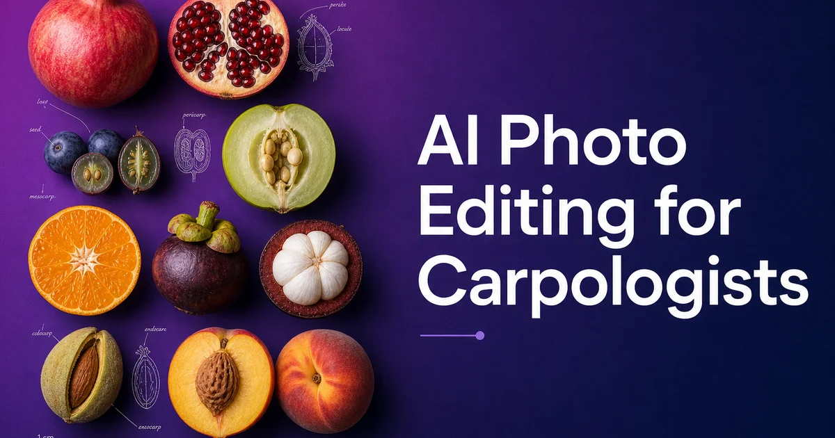
A carpologia — o estudo de frutos e sementes — ocupa uma interseção crítica entre botânica, agricultura e arqueologia. A paleobotânica depende de registros visuais detalhados para identificação de espécies, descrição taxonômica, pesquisa de evolução de culturas e interpretação de sítios arqueológicos. Sementes e frutos estão entre os restos vegetais mais comumente recuperados em escavações arqueológicas. Sua identificação fornece evidências diretas da dieta, agricultura, comércio e ambiente antigos. Na botânica e agricultura modernas, o estudo carpológico apoia a curadoria de bancos de sementes, programas de melhoramento de culturas, identificação de ervas daninhas e avaliação de biodiversidade. Em todas essas aplicações, a fotografia de alta qualidade de espécimes é fundamental para documentar as características morfológicas que possibilitam a identificação e comparação.
Os desafios fotográficos na carpologia surgem do pequeno tamanho da maioria dos espécimes, da natureza sutil das características superficiais diagnósticas e dos diversos estados de preservação encontrados no material de pesquisa. As sementes variam desde sementes de orquídea semelhantes a poeira com menos de um milímetro de comprimento até grandes frutos de palmeira. A grande maioria dos espécimes taxonomicamente importantes está na faixa de um a dez milímetros, exigindo macrofotografia com gerenciamento preciso de profundidade de campo. A escultura superficial — os padrões de fossas, cristas, reticulações, papilas e estriações no tegumento da semente — é frequentemente o caractere diagnóstico primário, mas essas características podem ter relevo de apenas dezenas de micrômetros e são facilmente perdidas em fotografias com iluminação imperfeita ou resolução insuficiente.
As ferramentas de edição de fotos com IA abordam diretamente esses desafios automatizando as etapas de pós-processamento que os carpologistas realizam em praticamente cada imagem de espécime. A remoção de fundo isola sementes e frutos da matriz de solo, bandejas de triagem e desordem do laboratório. O aprimoramento de detalhes recupera a fina escultura superficial que direciona a identificação — reticulação do tegumento, lenticelas da superfície do fruto, morfologia do hilo e anatomia da seção transversal. O processamento em lote padroniza imagens de sessões fotográficas prolongadas em que a iluminação mudou entre os espécimes. Para pesquisadores gerenciando coleções de milhares de espécimes destinados a bancos de dados de referência, guias de identificação ou pranchas de publicação, o processamento eficiente de imagens não é uma conveniência, mas uma necessidade prática.
- A remoção de fundo isola espécimes de sementes e frutos da matriz de solo, bandejas de coleta e superfícies de laboratório para imagens de publicação limpas e análise morfométrica.
- O aprimoramento com IA aguça a escultura superficial diagnosticamente crítica — reticulação do tegumento, pontuação, estriação, papilas e morfologia do hilo — que direciona a identificação carpológica.
- Magic Eraser remove marcas de pinças, resíduos de adesivo e detritos de preparação sem alterar as características morfológicas diagnósticas dos espécimes.
- O processamento em lote padroniza imagens de sessões fotográficas prolongadas em que iluminação, ampliação e configurações da câmera variaram entre os espécimes.
- Exportações prontas para publicação em 300 DPI com barras de escala calibradas atendem aos requisitos de periódicos para descrições taxonômicas e relatórios arqueobotânicos.
Desafios da macrofotografia e soluções de IA para documentação de sementes e frutos
O desafio fotográfico fundamental na carpologia é capturar detalhes diagnósticos em espécimes que frequentemente medem apenas poucos milímetros em sua maior dimensão. Na ampliação necessária para preencher o quadro da câmera com uma semente de dois milímetros, a profundidade de campo pode ser inferior a meio milímetro. Isso significa que a superfície dorsal está nítida enquanto as margens laterais estão completamente desfocadas, ou vice-versa. O empilhamento de foco resolve isso combinando múltiplas imagens focadas em diferentes planos, gerando grande número de quadros-fonte que devem ser precisamente alinhados e mesclados. Para uma sessão fotográfica documentando cinquenta espécimes com múltiplas vistas cada, o número total de quadros-fonte pode chegar a milhares.
O pós-processamento com IA se integra ao fluxo de trabalho de empilhamento de foco em múltiplos pontos. Após o empilhamento, o aprimoramento com IA aguça detalhes que o algoritmo de mesclagem não resolveu completamente, principalmente nos limites entre zonas focais onde um leve desalinhamento produz transições suaves. A remoção de fundo é especialmente valiosa para imagens empilhadas porque o empilhamento de foco frequentemente introduz artefatos de borda — halos brilhantes e imagens fantasma no limite do espécime onde os fundos desfocados de diferentes camadas do empilhamento foram combinados imperfeitamente. A remoção com IA elimina esses artefatos de forma limpa preservando a borda nítida do espécime que o empilhamento pretendia produzir.
A iluminação para fotografia carpológica requer cuidado particular porque as superfícies das sementes apresentam uma ampla gama de propriedades ópticas. Algumas sementes são altamente refletivas com tegumentos brilhantes que produzem reflexos especulares; outras são foscas com superfícies escuras que absorvem a luz. Alguns espécimes têm características superficiais definidas por sombras — fossas e cristas esculpidas visíveis apenas pelas sombras que projetam sob iluminação direcional — enquanto outros têm características definidas por diferenças de refletância. A normalização de exposição com IA e a recuperação de sombras ajudam a gerenciar esses extremos ópticos, produzindo imagens onde os detalhes superficiais são visíveis independentemente de o tegumento ser altamente refletivo ou profundamente absorvente.
- A profundidade de campo em macroampliação pode ser inferior a meio milímetro em uma semente de dois milímetros, exigindo empilhamento de foco que gera milhares de quadros-fonte em uma sessão típica.
- A remoção de fundo com IA elimina de forma limpa os artefatos de borda do empilhamento de foco — halos e imagens fantasma — preservando os limites nítidos do espécime produzidos pelo empilhamento.
- A recuperação de sombras e normalização de exposição lidam com a ampla gama de propriedades ópticas da superfície das sementes, desde tegumentos brilhantes altamente refletivos até superfícies escuras profundamente absorventes.
- O aprimoramento com IA pós-empilhamento aguça detalhes nos limites de zonas focais onde o algoritmo de mesclagem produziu transições suaves entre regiões nítidas.
Aprimoramento da escultura superficial diagnóstica para identificação taxonômica
A escultura do tegumento — o padrão superficial tridimensional do exterior da semente — é frequentemente o caractere diagnóstico mais importante para a identificação carpológica. A gama de tipos de superfície é enorme: superfícies reticuladas com uma rede de cristas elevadas encerrando células deprimidas, superfícies estriadas com cristas paralelas, superfícies papiladas cobertas com pequenas projeções arredondadas, superfícies pontuadas com depressões regular ou irregularmente espaçadas, superfícies verrucosas com protuberâncias semelhantes a verrugas e superfícies lisas que ainda mostram impressões celulares sob ampliação suficiente. Esses tipos de superfície e seus parâmetros específicos — tamanho celular, largura das cristas, profundidade das fossas, densidade de papilas — podem ser diagnósticos nos níveis de família, gênero e espécie.
O aprimoramento com IA aborda o desafio de tornar essas características superficiais sutis claramente visíveis em fotografias. Muitas esculturas do tegumento têm relevo medido em dezenas de micrômetros — raso o suficiente para aparecer como textura tênue em vez de padrão tridimensional claro em macrofotografias padrão. O aprimoramento de contraste local com IA aumenta seletivamente a visibilidade do relevo superficial amplificando as variações tonais em pequena escala causadas pela reflexão diferencial de luz das características superficiais elevadas e deprimidas. O resultado mostra a escultura superficial com a clareza que a microscopia eletrônica de varredura proporciona, mas em imagens coloridas com a aparência natural do espécime em vez da aparência artificial em escala de cinza das imagens de MEV.
A fotografia de seções transversais adiciona outra dimensão aos registros carpológicos. O corte de uma semente ou fruto para revelar a anatomia interna — forma e posição do embrião, presença e textura do endosperma, diferenciação das camadas do pericarpo e arranjo dos feixes vasculares — fornece informações diagnósticas não visíveis nas vistas externas. Seções transversais frequentemente apresentam superfícies irregulares onde a ferramenta de corte deixou marcas de rasgos e artefatos de compressão, principalmente em espécimes pequenos onde o corte de precisão é difícil. A limpeza com IA remove esses artefatos de preparação preservando os limites naturais dos tecidos e as estruturas celulares que são diagnosticamente importantes.
- A escultura do tegumento — reticulada, estriada, papilada, pontuada, verrucosa — fornece caracteres diagnósticos do nível de família ao de espécie com parâmetros medidos em escalas submilimétricas.
- O aprimoramento de contraste local com IA amplifica o relevo superficial raso medido em dezenas de micrômetros para mostrar a escultura com clareza de microscópio eletrônico de varredura em imagens coloridas.
- Artefatos de preparação de seções transversais causados por ferramentas de corte — marcas de rasgo, danos por compressão — são removidos pela limpeza com IA preservando limites diagnósticos de tecidos e estruturas celulares.
- A combinação de imagens de superfície externa aprimoradas e seções transversais limpas fornece documentação morfológica abrangente para fins taxonômicos e de identificação.
Aplicações arqueobotânicas: documentação de espécimes carbonizados, encharcados e mineralizados
A arqueobotânica — o estudo de restos vegetais de sítios arqueológicos — depende fortemente da identificação carpológica de sementes e frutos recuperados de escavações. Esses espécimes foram preservados por carbonização (queima em incêndios antigos), encharcamento (submersão em depósitos anaeróbicos saturados de água) ou mineralização (substituição do tecido orgânico por fosfato de cálcio em depósitos de latrina ou lixeira). Cada via de preservação altera a morfologia do espécime original de maneiras específicas e as fotografias devem documentar tanto o estado de preservação quanto as características diagnósticas que sobreviveram à alteração. A edição de fotos com IA é especialmente valiosa para o trabalho arqueobotânico porque os espécimes são frequentemente frágeis, danificados e contaminados com sedimento aderente.
Sementes carbonizadas — de longe o achado arqueobotânico mais comum — foram reduzidas a carbono puro por incêndios antigos, encolhendo em tamanho e frequentemente distorcendo em forma. A escultura superficial original pode estar parcialmente preservada, mas agora é renderizada inteiramente em preto, tornando muito difícil fotografá-la com contraste suficiente para mostrar detalhes superficiais. O aprimoramento de contraste com IA é transformador para a fotografia de espécimes carbonizados, recuperando a escultura superficial da superfície quase uniformemente preta ao amplificar as minúsculas diferenças de refletância entre características superficiais elevadas e deprimidas. Esse aprimoramento pode tornar características diagnósticas visíveis em fotografias que, de outra forma, apareceriam como formas pretas sem detalhes.
Sementes encharcadas retêm seu tecido orgânico original, mas estão amolecidas e frágeis pela submersão prolongada. Devem ser fotografadas enquanto ainda estão molhadas — a secagem causa encolhimento e distorção irreversíveis — o que introduz reflexos, filmes de água superficiais e partículas de sedimento aderentes. Espécimes mineralizados podem reter detalhes superficiais notáveis, mas frequentemente têm uma cor mineral uniforme que obscurece a variação natural de cor entre diferentes tecidos. Em cada caso, a edição com IA aborda os desafios fotográficos específicos do tipo de preservação: recuperação de contraste para material carbonizado, remoção de reflexos e limpeza de sedimentos para material encharcado e aprimoramento de diferenciação de tecidos para espécimes mineralizados.
- Sementes carbonizadas reduzidas a carbono preto uniforme se beneficiam do aprimoramento de contraste com IA que recupera a escultura superficial de minúsculas diferenças de refletância invisíveis em fotografias padrão.
- Espécimes encharcados fotografados molhados requerem remoção com IA de reflexos superficiais, filmes de água e partículas de sedimento aderentes enquanto o material frágil permanece hidratado.
- Espécimes mineralizados com coloração mineral uniforme se beneficiam do aprimoramento com IA que diferencia tipos de tecido com base em variações sutis de densidade e textura.
- Cada via de preservação introduz desafios fotográficos específicos que a edição com IA aborda com estratégias de aprimoramento direcionadas correspondentes ao tipo de preservação.
Análise morfométrica e bancos de dados digitais de referência de sementes
A carpologia moderna depende cada vez mais da análise morfométrica — medição quantitativa da forma, tamanho e características superficiais de sementes e frutos — tanto para identificação quanto para pesquisa evolutiva. Software de medição automatizado extrai parâmetros de fotografias de espécimes incluindo comprimento, largura, espessura, área, perímetro, circularidade, índice de elongação e descritores de textura superficial. A precisão dessas medições depende diretamente da qualidade da imagem: fundos limpos para detecção precisa de contorno, foco nítido para delineação precisa de limites e referências de escala calibradas para precisão dimensional absoluta. A edição de fotos com IA produz imagens otimizadas para análise morfométrica garantindo espécimes limpos em fundos uniformes com bordas nítidas.
Bancos de dados digitais de referência de sementes — coleções de imagens padronizadas usadas para identificação por comparação — atendem tanto aplicações botânicas modernas quanto arqueobotânicas. O Millennium Seed Bank em Kew, o banco de dados GRIN do USDA e muitos bancos de dados de herbários regionais mantêm coleções crescentes de imagens de sementes para referência de identificação. Contribuir com imagens de alta qualidade para esses bancos de dados requer protocolos fotográficos padronizados e pós-processamento consistente para garantir que imagens de diferentes contribuidores mantenham coerência visual. O processamento em lote com IA com parâmetros de aprimoramento consistentes normaliza imagens de fontes diversas em uma coleção de referência visualmente unificada.
Abordagens de aprendizado de máquina para identificação automatizada de sementes são uma aplicação emergente que depende de grandes conjuntos de dados de imagens de sementes de alta qualidade. Treinar classificadores de imagem para identificar espécies a partir de fotografias requer milhares de imagens rotuladas por espécie com fundos consistentes, orientações padronizadas e características diagnósticas claramente visíveis. Ferramentas de edição de fotos com IA aceleram a produção de conjuntos de dados de treinamento processando em lote fotografias brutas de coleções no formato padronizado que algoritmos de aprendizado de máquina requerem. À medida que sistemas de identificação automatizada amadurecem, a qualidade de seus dados de treinamento — e, portanto, a qualidade das imagens-fonte e do pós-processamento — determina diretamente a precisão das identificações que produzem.
- Software de análise morfométrica requer fundos limpos para detecção de contorno, foco nítido para delineação de limites e escalas calibradas — tudo aprimorado pelo processamento de imagem com IA.
- Bancos de dados digitais de referência de sementes mantidos por instituições como Kew e USDA se beneficiam da normalização em lote com IA que cria coerência visual entre contribuições de fontes diversas.
- Conjuntos de dados de treinamento para identificação de sementes por aprendizado de máquina requerem milhares de imagens padronizadas por espécie que o processamento em lote com IA produz eficientemente a partir de fotografias brutas de coleções.
- A precisão dos sistemas emergentes de identificação automatizada depende diretamente da qualidade das imagens de treinamento, tornando o pós-processamento com IA um investimento fundamental para a futura tecnologia carpológica.
How to do it
- 1
Photograph fruit and seed specimens with standardized macro setups and calibrated scale references
Set up a macro photography station with diffused lighting from multiple angles to minimize harsh shadows on seed and fruit surfaces. Include a millimeter scale bar in every frame for morphometric calibration. Photograph specimens from multiple standardized views — dorsal, ventral, lateral, and cross-section for seeds. Whole fruit, longitudinal section, and transverse section for fruits. Use a consistent neutral background and focus stacking for specimens where full-depth sharpness is needed to document surface sculpture and morphological details.
- 2
Remove distracting backgrounds and isolate specimens for publication and analysis
Use Background Eraser to isolate seed and fruit specimens from collection trays, soil matrix, sorting dishes, and laboratory surfaces. Archaeobotanical specimens recovered from excavations often retain adhering soil particles, root fragments, and mineral deposits that obscure surface features. Modern comparative specimens may be photographed on herbarium paper, in petri dishes, or on sorting mats with visible grid lines. Clean isolation against a uniform background is key for both publication plates and morphometric analysis software that requires clean specimen outlines.
- 3
Enhance fine surface sculpture and morphological details critical for identification
Use AI Enhance to sharpen the surface details that drive carpological identification. Seed coat sculpture including pitting, reticulation, striation, and papillae. Fruit surface features including lenticels, trichomes, and wax bloom texture. Hilum and micropyle morphology. And the internal anatomy visible in cross-sections including embryo position, endosperm structure, and pericarp layer differentiation. Many of these features are measured at sub-millimeter scale and require maximum image clarity for accurate records.
- 4
Clean up preparation artifacts and specimen damage
Use Magic Eraser to remove preparation artifacts. Forceps marks, dissection debris, mounting adhesive residue, and minor specimen damage — without altering diagnostic morphological features. This is mainly valuable for archaeobotanical specimens where carbonization, waterlogging, or mineralization has altered the specimen surface. Modern preparation handling may have introduced extra damage that should not be documented as original morphology.
- 5
Prepare multi-view figure plates and morphometric image sets
Arrange standardized multi-view images into publication-ready figure plates with consistent magnification, spacing, scale bars, and labels. Export at 300 DPI minimum for journal submission. For morphometric studies, prepare extra images with clean backgrounds and calibrated scale references optimized for automated measurement software that extracts dimensions, area, perimeter, circularity. Surface texture parameters from specimen photographs.
Fontes
- Standardized Photography Protocols for Seed and Fruit Morphology — Royal Botanic Gardens, Kew — Millennium Seed Bank
- Digital Imaging Techniques for Archaeobotanical Remains — Vegetation History and Archaeobotany — Springer
- Morphometric Analysis of Seeds Using Image Processing — Computers and Electronics in Agriculture — Elsevier